7LYU

Reelin repeat 8


Experimental Data Snapshot

  • Method: X-RAY DIFFRACTION
  • Resolution: 3.00 Å
  • R-Value Free: 0.264 
  • R-Value Work: 0.227 
  • R-Value Observed: 0.229 

Starting Model: experimental
View more details

wwPDB Validation   3D Report Full Report


This is version 1.2 of the entry. See complete history


Literature

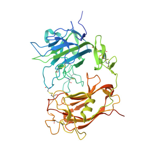
Structure of Reelin repeat 8 and the adjacent C-terminal region.

Turk, L.S.Currie, M.J.Dobson, R.C.J.Comoletti, D.

(2022) Biophys J 121: 2526-2537

  • DOI: https://doi.org/10.1016/j.bpj.2022.06.002
  • Primary Citation of Related Structures:  
    7LYU

  • PubMed Abstract: 

    Neuronal development and function are dependent in part on the several roles of the secreted glycoprotein Reelin. Endogenous proteases process this 400 kDa, modular protein, yielding N-terminal, central, and C-terminal fragments that each have distinct roles in Reelin's function and regulation. The C-terminal fragment comprises Reelin repeat (RR) domains seven and eight, as well as a basic stretch of 32 amino acid residues termed the C-terminal region (CTR), influences Reelin signaling intensity, and has been reported to bind to Neuropilin-1, which serves as a co-receptor in the canonical Reelin signaling pathway. Here, we present a crystal structure of RR8 at 3.0 Å resolution. Analytical ultracentrifugation and small-angle x-ray scattering confirmed that RR8 is monomeric and enabled us to identify the CTR as a flexible, yet compact subdomain. We conducted structurally informed protein engineering to design a chimeric RR8 construct guided by the structural similarities with RR6. Experimental results support a mode of Reelin-receptor interaction reliant on the multiple interfaces coordinating the binding event. Structurally, RR8 resembles other individual RRs, but its structure does show discrete differences that may account for Reelin receptor specificity toward RR6.


  • Organizational Affiliation

    Child Health Institute of New Jersey, New Brunswick, New Jersey; Department of Neuroscience and Cell Biology, Robert Wood Johnson Medical School, Rutgers, The State University of New Jersey, New Brunswick, New Jersey; School of Biological Sciences, Victoria University of Wellington, Wellington, New Zealand. Electronic address: liam.turk@otago.ac.nz.


Macromolecules
Find similar proteins by:  (by identity cutoff)  |  3D Structure
Entity ID: 1
MoleculeChains Sequence LengthOrganismDetailsImage
Reelin
A, B
414Mus musculusMutation(s): 0 
Gene Names: RelnRl
EC: 3.4.21
UniProt & NIH Common Fund Data Resources
Find proteins for Q60841 (Mus musculus)
Explore Q60841 
Go to UniProtKB:  Q60841
IMPC:  MGI:103022
Entity Groups  
Sequence Clusters30% Identity50% Identity70% Identity90% Identity95% Identity100% Identity
UniProt GroupQ60841
Glycosylation
Glycosylation Sites: 2Go to GlyGen: Q60841-1
Sequence Annotations
Expand
  • Reference Sequence
Oligosaccharides

Help

Entity ID: 2
MoleculeChains Length2D Diagram Glycosylation3D Interactions
alpha-D-mannopyranose-(1-3)-alpha-D-mannopyranose-(1-6)-[alpha-D-mannopyranose-(1-3)]beta-D-mannopyranose-(1-4)-2-acetamido-2-deoxy-beta-D-glucopyranose-(1-4)-2-acetamido-2-deoxy-beta-D-glucopyranose
C
6N-Glycosylation
Glycosylation Resources
GlyTouCan:  G09724ZC
GlyCosmos:  G09724ZC
GlyGen:  G09724ZC
Entity ID: 3
MoleculeChains Length2D Diagram Glycosylation3D Interactions
alpha-D-mannopyranose-(1-6)-beta-D-mannopyranose-(1-4)-2-acetamido-2-deoxy-beta-D-glucopyranose-(1-4)-2-acetamido-2-deoxy-beta-D-glucopyranose
D
4N-Glycosylation
Glycosylation Resources
GlyTouCan:  G22573RC
GlyCosmos:  G22573RC
GlyGen:  G22573RC
Entity ID: 4
MoleculeChains Length2D Diagram Glycosylation3D Interactions
alpha-D-mannopyranose-(1-2)-alpha-D-mannopyranose-(1-3)-beta-D-mannopyranose-(1-4)-2-acetamido-2-deoxy-beta-D-glucopyranose-(1-4)-2-acetamido-2-deoxy-beta-D-glucopyranose
E
5N-Glycosylation
Glycosylation Resources
GlyTouCan:  G42227JK
GlyCosmos:  G42227JK
GlyGen:  G42227JK
Entity ID: 5
MoleculeChains Length2D Diagram Glycosylation3D Interactions
beta-D-mannopyranose-(1-4)-2-acetamido-2-deoxy-beta-D-glucopyranose-(1-4)-2-acetamido-2-deoxy-beta-D-glucopyranose
F
3N-Glycosylation
Glycosylation Resources
GlyTouCan:  G15407YE
GlyCosmos:  G15407YE
GlyGen:  G15407YE
Experimental Data & Validation

Experimental Data

  • Method: X-RAY DIFFRACTION
  • Resolution: 3.00 Å
  • R-Value Free: 0.264 
  • R-Value Work: 0.227 
  • R-Value Observed: 0.229 
  • Space Group: P 1 21 1
Unit Cell:
Length ( Å )Angle ( ˚ )
a = 60.171α = 90
b = 104.059β = 104.109
c = 67.615γ = 90
Software Package:
Software NamePurpose
PHENIXrefinement
XDSdata reduction
Aimlessdata scaling
MrBUMPphasing

Structure Validation

View Full Validation Report


Entry History & Funding Information

Deposition Data


Funding OrganizationLocationGrant Number
National Science Foundation (NSF, United States)United States1755189

Revision History  (Full details and data files)

  • Version 1.0: 2022-06-15
    Type: Initial release
  • Version 1.1: 2022-12-28
    Changes: Database references, Structure summary
  • Version 1.2: 2023-10-25
    Changes: Data collection, Refinement description