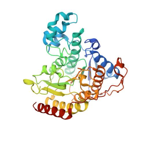
7LTL

STRUCTURE OF HUMAN HDAC2 IN COMPLEX WITH AN INHIBITOR LACKING A ZINC BINDING GROUP (COMPOUND 19)


Experimental Data Snapshot

  • Method: X-RAY DIFFRACTION
  • Resolution: 1.49 Å
  • R-Value Free: 0.195 
  • R-Value Work: 0.170 
  • R-Value Observed: 0.171 

wwPDB Validation   3D Report Full Report


Ligand Structure Quality Assessment 


This is version 1.1 of the entry. See complete history


Literature

Redefining the Histone Deacetylase Inhibitor Pharmacophore: High Potency with No Zinc Cofactor Interaction.

Beshore, D.C.Adam, G.C.Barnard, R.J.O.Burlein, C.Gallicchio, S.N.Holloway, M.K.Krosky, D.Lemaire, W.Myers, R.W.Patel, S.Plotkin, M.A.Powell, D.A.Rada, V.Cox, C.D.Coleman, P.J.Klein, D.J.Wolkenberg, S.E.

(2021) ACS Med Chem Lett 12: 540-547

  • DOI: https://doi.org/10.1021/acsmedchemlett.1c00074
  • Primary Citation of Related Structures:  
    7LTG, 7LTK, 7LTL

  • PubMed Abstract: 

    A novel series of histone deacetylase (HDAC) inhibitors lacking a zinc-binding moiety has been developed and described herein. HDAC isozyme profiling and kinetic studies indicate that these inhibitors display a selectivity preference for HDACs 1, 2, 3, 10, and 11 via a rapid equilibrium mechanism, and crystal structures with HDAC2 confirm that these inhibitors do not interact with the catalytic zinc. The compounds are nonmutagenic and devoid of electrophilic and mutagenic structural elements and exhibit off-target profiles that are promising for further optimization. The efficacy of this new class in biochemical and cell-based assays is comparable to the marketed HDAC inhibitors belinostat and vorinostat. These results demonstrate that the long-standing pharmacophore model of HDAC inhibitors requiring a metal binding motif should be revised and offers a distinct class of HDAC inhibitors.


  • Organizational Affiliation

    MRL, Merck & Co., Inc., Kenilworth, New Jersey 07033, United States.


Macromolecules
Find similar proteins by:  (by identity cutoff)  |  3D Structure
Entity ID: 1
MoleculeChains Sequence LengthOrganismDetailsImage
Histone deacetylase 2
A, B, C
376Homo sapiensMutation(s): 0 
Gene Names: HDAC2
EC: 3.5.1.98 (PDB Primary Data), 3.5.1 (UniProt)
UniProt & NIH Common Fund Data Resources
Find proteins for Q92769 (Homo sapiens)
Explore Q92769 
Go to UniProtKB:  Q92769
PHAROS:  Q92769
GTEx:  ENSG00000196591 
Entity Groups  
Sequence Clusters30% Identity50% Identity70% Identity90% Identity95% Identity100% Identity
UniProt GroupQ92769
Sequence Annotations
Expand
  • Reference Sequence
Small Molecules
Ligands 6 Unique
IDChains Name / Formula / InChI Key2D Diagram3D Interactions
YEV (Subject of Investigation/LOI)
Query on YEV

Download Ideal Coordinates CCD File 
JA [auth C],
O [auth A],
Y [auth B]
(2R)-2-(5-hydroxy-2-methyl-1H-indol-3-yl)-N-{(1S)-1-[5-(2-methoxyquinolin-3-yl)-1H-imidazol-2-yl]pentyl}propanamide
C30 H33 N5 O3
CZYALTGXIQQGCT-NSYGIPOTSA-N
PEG
Query on PEG

Download Ideal Coordinates CCD File 
IA [auth C]
L [auth A]
M [auth A]
N [auth A]
W [auth B]
IA [auth C],
L [auth A],
M [auth A],
N [auth A],
W [auth B],
X [auth B]
DI(HYDROXYETHYL)ETHER
C4 H10 O3
MTHSVFCYNBDYFN-UHFFFAOYSA-N
SO4
Query on SO4

Download Ideal Coordinates CCD File 
AA [auth C]
BA [auth C]
CA [auth C]
DA [auth C]
E [auth A]
AA [auth C],
BA [auth C],
CA [auth C],
DA [auth C],
E [auth A],
EA [auth C],
F [auth A],
G [auth A],
H [auth A],
Q [auth B],
R [auth B],
S [auth B]
SULFATE ION
O4 S
QAOWNCQODCNURD-UHFFFAOYSA-L
ZN
Query on ZN

Download Ideal Coordinates CCD File 
D [auth A],
P [auth B],
Z [auth C]
ZINC ION
Zn
PTFCDOFLOPIGGS-UHFFFAOYSA-N
ACT
Query on ACT

Download Ideal Coordinates CCD File 
HA [auth C],
K [auth A],
V [auth B]
ACETATE ION
C2 H3 O2
QTBSBXVTEAMEQO-UHFFFAOYSA-M
CA
Query on CA

Download Ideal Coordinates CCD File 
FA [auth C]
GA [auth C]
I [auth A]
J [auth A]
T [auth B]
FA [auth C],
GA [auth C],
I [auth A],
J [auth A],
T [auth B],
U [auth B]
CALCIUM ION
Ca
BHPQYMZQTOCNFJ-UHFFFAOYSA-N
Binding Affinity Annotations 
IDSourceBinding Affinity
YEV BindingDB:  7LTL IC50: min: 37, max: 3200 (nM) from 5 assay(s)
Experimental Data & Validation

Experimental Data

  • Method: X-RAY DIFFRACTION
  • Resolution: 1.49 Å
  • R-Value Free: 0.195 
  • R-Value Work: 0.170 
  • R-Value Observed: 0.171 
  • Space Group: P 21 21 21
Unit Cell:
Length ( Å )Angle ( ˚ )
a = 92.35α = 90
b = 98.76β = 90
c = 139.44γ = 90
Software Package:
Software NamePurpose
BUSTERrefinement
PDB_EXTRACTdata extraction
XDSdata reduction
Aimlessdata scaling
BUSTERphasing

Structure Validation

View Full Validation Report


Ligand Structure Quality Assessment 


Entry History 

Deposition Data

Revision History  (Full details and data files)

  • Version 1.0: 2021-05-05
    Type: Initial release
  • Version 1.1: 2024-03-06
    Changes: Data collection, Database references