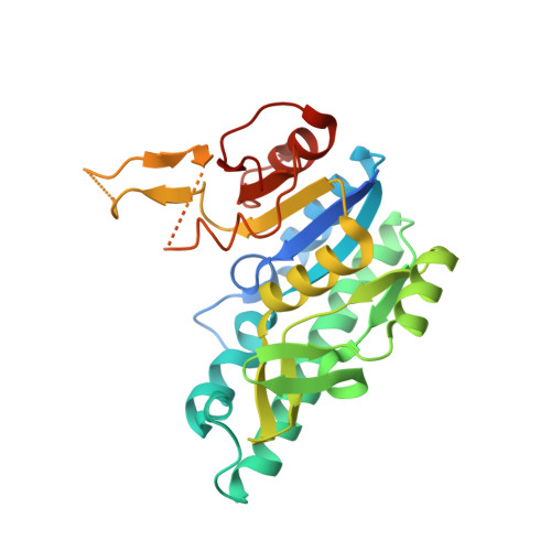
Molecular Basis for the Substrate Promiscuity of Isopentenyl Phosphate Kinase from Candidatus methanomethylophilus alvus .
Johnson, B.P., Kumar, V., Scull, E.M., Thomas, L.M., Bourne, C.R., Singh, S.(2022) ACS Chem Biol 17: 85-102
- PubMed: 34905349
- DOI: https://doi.org/10.1021/acschembio.1c00655
- Primary Citation of Related Structures:
7LNT, 7LNU, 7LNV, 7LNW, 7LNX, 7N9D - PubMed Abstract:
Isopentenyl phosphate kinases (IPKs) catalyze the ATP-dependent phosphorylation of isopentenyl monophosphate (IP) to isopentenyl diphosphate (IPP) in the alternate mevalonate pathways of the archaea and plant cytoplasm. In recent years, IPKs have also been employed in artificial biosynthetic pathways called "(iso) prenol pathways" that utilize promiscuous kinases to sequentially phosphorylate (iso) prenol and generate the isoprenoid precursors IPP and dimethylallyl diphosphate (DMAPP). Furthermore, IPKs have garnered attention for their impressive substrate promiscuity toward non-natural alkyl-monophosphates (alkyl-Ps), which has prompted their utilization as biocatalysts for the generation of novel isoprenoids. However, none of the IPK crystal structures currently available contain non-natural substrates, leaving the roles of active-site residues in substrate promiscuity ambiguous. To address this, we present herein the high-resolution crystal structures of an IPK from Candidatus methanomethylophilus alvus (CMA) in the apo form and bound to natural and non-natural substrates. Additionally, we describe active-site engineering studies leading to enzyme variants with broadened substrate scope, as well as structure determination of two such variants (Ile74Ala and Ile146Ala) bound to non-natural alkyl-Ps. Collectively, our crystallographic studies compare six structures of CMA variants in different ligand-bound forms and highlight contrasting structural dynamics of the two substrate-binding sites. Furthermore, the structural and mutational studies confirm a novel role of the highly conserved DVTGG motif in catalysis, both in CMA and in IPKs at large. As such, the current study provides a molecular basis for the substrate-binding modes and catalytic performance of CMA toward the goal of developing IPKs into useful biocatalysts.
- Department of Chemistry and Biochemistry, University of Oklahoma, Stephenson Life Sciences Research Center, 101 Stephenson Pkwy, Norman, Oklahoma 73019, United States.
Organizational Affiliation:


