Turnover and Inactivation Mechanisms for ( S )-3-Amino-4,4-difluorocyclopent-1-enecarboxylic Acid, a Selective Mechanism-Based Inactivator of Human Ornithine Aminotransferase.
Shen, S., Butrin, A., Doubleday, P.F., Melani, R.D., Beaupre, B.A., Tavares, M.T., Ferreira, G.M., Kelleher, N.L., Moran, G.R., Liu, D., Silverman, R.B.(2021) J Am Chem Soc 143: 8689-8703
- PubMed: 34097381 Search on PubMedSearch on PubMed Central
- DOI: https://doi.org/10.1021/jacs.1c02456
- Primary Citation Related Structures:
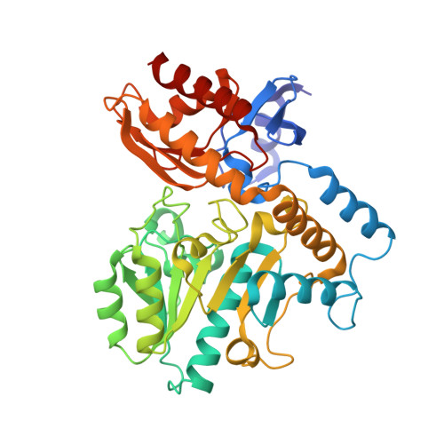
7LK0, 7LK1 - PubMed Abstract:
The inhibition of human ornithine δ-aminotransferase ( h OAT) is a potential therapeutic approach to treat hepatocellular carcinoma. In this work, ( S )-3-amino-4,4-difluorocyclopent-1-enecarboxylic acid (SS-1-148, 6 ) was identified as a potent mechanism-based inactivator of h OAT while showing excellent selectivity over other related aminotransferases (e.g., GABA-AT). An integrated mechanistic study was performed to investigate the turnover and inactivation mechanisms of 6 . A monofluorinated ketone ( M10 ) was identified as the primary metabolite of 6 in h OAT. By soaking h OAT holoenzyme crystals with 6 , a precursor to M10 was successfully captured. This gem -diamine intermediate, covalently bound to Lys292, observed for the first time in h OAT/ligand crystals, validates the turnover mechanism proposed for 6 . Co-crystallization yielded h OAT in complex with 6 and revealed a novel noncovalent inactivation mechanism in h OAT. Native protein mass spectrometry was utilized for the first time in a study of an aminotransferase inactivator to validate the noncovalent interactions between the ligand and the enzyme; a covalently bonded complex was also identified as a minor form observed in the denaturing intact protein mass spectrum. Spectral and stopped-flow kinetic experiments supported a lysine-assisted E2 fluoride ion elimination, which has never been observed experimentally in other studies of related aminotransferase inactivators. This elimination generated the second external aldimine directly from the initial external aldimine, rather than the typical E1cB elimination mechanism, forming a quinonoid transient state between the two external aldimines. The use of native protein mass spectrometry, X-ray crystallography employing both soaking and co-crystallization methods, and stopped-flow kinetics allowed for the detailed elucidation of unusual turnover and inactivation pathways.
- Department of Chemistry, Center for Molecular Innovation and Drug Discovery, and Center for Developmental Therapeutics, Northwestern University, Evanston, Illinois 60208, United States.
Organizational Affiliation: