Photoisomerization detected in a fully wavelength-tunable rhodopsin mimic system.
Ehyaei, N., Bingham, C., Silva, K., Nossoni, Z., Gavgani, H.N., Nosrati, M., Eaves, J., Akhdar, M., Vasileiou, C., Borhan, B., Geiger, J.H.(2026) Acta Crystallogr D Struct Biol 82: 664-671
- PubMed: 42201784 Search on PubMedSearch on PubMed Central
- DOI: https://doi.org/10.1107/S2059798326003839
- Primary Citation Related Structures:
7LHM, 7LHN, 7LHO, 9PN1 - PubMed Abstract:
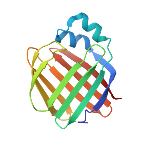
We describe the photoisomerization of the retinylidene protonated Schiff base in human retinol-binding protein II (hCRBPII) and the role of water molecules in this process. We characterize the photoisomerization of the 15-cis/all-trans retinylidene protonated Schiff base in this system using UV-visible spectroscopy and atomic-resolution X-ray crystallography. We further demonstrate a process where the pK a of the protonated Schiff base is substantially altered by light-induced dehydration of the binding pocket, suggesting novel pathways of photoswitching that rely not on isomerization or conformational change of the chromophore but rather on light-induced reorganization of the protein environment.
- Department of Chemistry, Michigan State University, East Lansing, MI 48824, USA.
Organizational Affiliation: