7L6W | pdb_00007l6w

SFX structure of the MyD88 TIR domain higher-order assembly


Experimental Data Snapshot

  • Method: X-RAY DIFFRACTION
  • Resolution: 2.30 Å
  • R-Value Free: 
    0.288 (Depositor), 0.277 (DCC) 
  • R-Value Work: 
    0.233 (Depositor), 0.224 (DCC) 
  • R-Value Observed: 
    0.238 (Depositor) 

Starting Model: experimental
View more details

wwPDB Validation 3D Report Full Report

Validation slider image for 7L6W

This is version 1.3 of the entry. See complete history

Literature

MyD88 TIR domain higher-order assembly interactions revealed by microcrystal electron diffraction and serial femtosecond crystallography.

Clabbers, M.T.B.Holmes, S.Muusse, T.W.Vajjhala, P.R.Thygesen, S.J.Malde, A.K.Hunter, D.J.B.Croll, T.I.Flueckiger, L.Nanson, J.D.Rahaman, M.H.Aquila, A.Hunter, M.S.Liang, M.Yoon, C.H.Zhao, J.Zatsepin, N.A.Abbey, B.Sierecki, E.Gambin, Y.Stacey, K.J.Darmanin, C.Kobe, B.Xu, H.Ve, T.

(2021) Nat Commun 12: 2578-2578

  • DOI: https://doi.org/10.1038/s41467-021-22590-6
  • Primary Citation Related Structures: 
    7BEQ, 7BER, 7L6W

  • PubMed Abstract: 

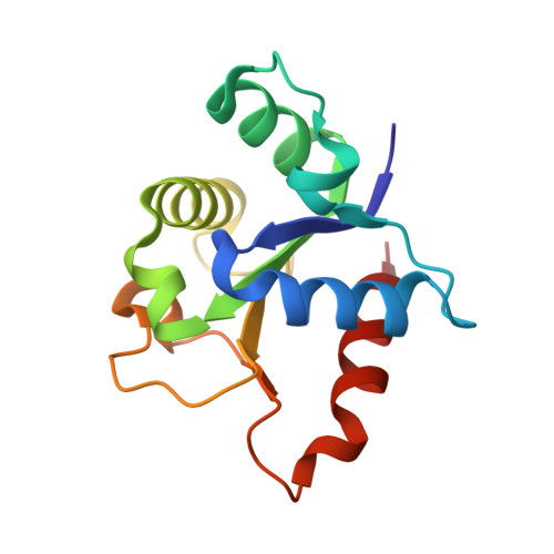
    MyD88 and MAL are Toll-like receptor (TLR) adaptors that signal to induce pro-inflammatory cytokine production. We previously observed that the TIR domain of MAL (MAL TIR ) forms filaments in vitro and induces formation of crystalline higher-order assemblies of the MyD88 TIR domain (MyD88 TIR ). These crystals are too small for conventional X-ray crystallography, but are ideally suited to structure determination by microcrystal electron diffraction (MicroED) and serial femtosecond crystallography (SFX). Here, we present MicroED and SFX structures of the MyD88 TIR assembly, which reveal a two-stranded higher-order assembly arrangement of TIR domains analogous to that seen previously for MAL TIR . We demonstrate via mutagenesis that the MyD88 TIR assembly interfaces are critical for TLR4 signaling in vivo, and we show that MAL promotes unidirectional assembly of MyD88 TIR . Collectively, our studies provide structural and mechanistic insight into TLR signal transduction and allow a direct comparison of the MicroED and SFX techniques.


  • Organizational Affiliation
    • Department of Materials and Environmental Chemistry, Stockholm University, Stockholm, Sweden.

Macromolecule Content 

  • Total Structure Weight: 16.21 kDa 
  • Atom Count: 1,183 
  • Modeled Residue Count: 138 
  • Deposited Residue Count: 138 
  • Unique protein chains: 1

Macromolecules

Find similar proteins by:|  3D Structure
Entity ID: 1
MoleculeChains  Sequence LengthOrganismDetailsImage
Myeloid differentiation primary response protein MyD88138Homo sapiensMutation(s): 0 
Gene Names: MYD88
UniProt & NIH Common Fund Data Resources
Find proteins for Q99836 (Homo sapiens)
Explore Q99836 
Go to UniProtKB:  Q99836
PHAROS:  Q99836
GTEx:  ENSG00000172936 
Entity Groups
Sequence Clusters30% Identity50% Identity70% Identity90% Identity95% Identity100% Identity
UniProt GroupQ99836
Sequence Annotations
Expand
Reference Sequence

Experimental Data & Validation

Experimental Data

  • Method: X-RAY DIFFRACTION
  • Resolution: 2.30 Å
  • R-Value Free:  0.288 (Depositor), 0.277 (DCC) 
  • R-Value Work:  0.233 (Depositor), 0.224 (DCC) 
  • R-Value Observed: 0.238 (Depositor) 
Space Group: C 1 2 1
Unit Cell:
Length ( Å )Angle ( ˚ )
a = 100.4α = 90
b = 31.5β = 107.4
c = 54.5γ = 90
Software Package:
Software NamePurpose
Aimlessdata scaling
REFMACrefinement
REFMACphasing
Aimlessdata reduction

Structure Validation

View Full Validation Report


Entry History 

& Funding Information

Deposition Data


Funding OrganizationLocationGrant Number
Australian Research Council (ARC)AustraliaCE140100011

Revision History  (Full details and data files)

  • Version 1.0: 2021-03-10
    Type: Initial release
  • Version 1.1: 2021-05-26
    Changes: Database references
  • Version 1.2: 2023-08-16
    Changes: Data collection, Database references
  • Version 1.3: 2023-10-18
    Changes: Refinement description