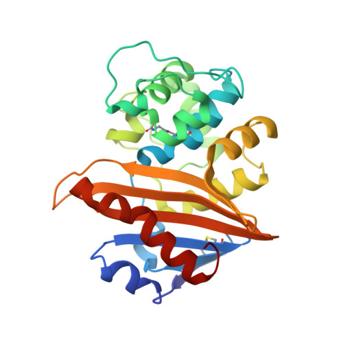
Crystal Structure of the Oxacillin-hydrolyzing Class D Extended-spectrum Beta-lactamase OXA-14 from Pseudomonas aeruginosa in Complex with Covalently Bound Clavulanic Acid
Minasov, G., Shuvalova, L., Rosas-Lemus, M., Brunzelle, J.S., Satchell, K.J.F., Center for Structural Biology of Infectious Diseases (CSBID)To be published.