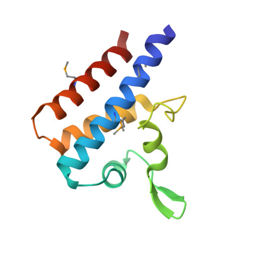
The ChiS-Family DNA-Binding Domain Contains a Cryptic Helix-Turn-Helix Variant.
Klancher, C.A., Minasov, G., Podicheti, R., Rusch, D.B., Dalia, T.N., Satchell, K.J.F., Neiditch, M.B., Dalia, A.B.(2021) mBio 12
- PubMed: 33727356 Search on PubMedSearch on PubMed Central
- DOI: https://doi.org/10.1128/mBio.03287-20
- Primary Citation Related Structures:
7KPO - PubMed Abstract:
Sequence-specific DNA-binding domains (DBDs) are conserved in all domains of life. These proteins carry out a variety of cellular functions, and there are a number of distinct structural domains already described that allow for sequence-specific DNA binding, including the ubiquitous helix-turn-helix (HTH) domain. In the facultative pathogen Vibrio cholerae , the chitin sensor ChiS is a transcriptional regulator that is critical for the survival of this organism in its marine reservoir. We recently showed that ChiS contains a cryptic DBD in its C terminus. This domain is not homologous to any known DBD, but it is a conserved domain present in other bacterial proteins. Here, we present the crystal structure of the ChiS DBD at a resolution of 1.28 Å. We find that the ChiS DBD contains an HTH domain that is structurally similar to those found in other DNA-binding proteins, like the LacI repressor. However, one striking difference observed in the ChiS DBD is that the canonical tight turn of the HTH is replaced with an insertion containing a β-sheet, a variant which we term the helix-sheet-helix. Through systematic mutagenesis of all positively charged residues within the ChiS DBD, we show that residues within and proximal to the ChiS helix-sheet-helix are critical for DNA binding. Finally, through phylogenetic analyses we show that the ChiS DBD is found in diverse proteobacterial proteins that exhibit distinct domain architectures. Together, these results suggest that the structure described here represents the prototypical member of the ChiS-family of DBDs. IMPORTANCE Regulating gene expression is essential in all domains of life. This process is commonly facilitated by the activity of DNA-binding transcription factors. There are diverse structural domains that allow proteins to bind to specific DNA sequences. The structural basis underlying how some proteins bind to DNA, however, remains unclear. Previously, we showed that in the major human pathogen Vibrio cholerae , the transcription factor ChiS directly regulates gene expression through a cryptic DNA-binding domain. This domain lacked homology to any known DNA-binding protein. In the current study, we determined the structure of the ChiS DNA-binding domain (DBD) and found that the ChiS-family DBD is a cryptic variant of the ubiquitous helix-turn-helix (HTH) domain. We further demonstrate that this domain is conserved in diverse proteins that may represent a novel group of transcriptional regulators.
- Department of Biology, Indiana University, Bloomington, Indiana, USA.
Organizational Affiliation: