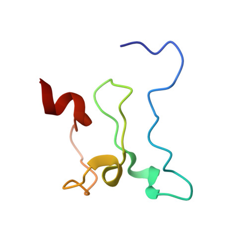
Recognition of Histone H3 Methylation States by the PHD1 Domain of Histone Demethylase KDM5A.
Longbotham, J.E., Kelly, M.J.S., Fujimori, D.G.(2021) ACS Chem Biol
- PubMed: 33621062
- DOI: https://doi.org/10.1021/acschembio.0c00976
- Primary Citation of Related Structures:
7KLO, 7KLR - PubMed Abstract:
PHD reader domains are chromatin binding modules often responsible for the recruitment of large protein complexes that contain histone modifying enzymes, chromatin remodelers, and DNA repair machinery. A majority of PHD domains recognize N-terminal residues of histone H3 and are sensitive to the methylation state of Lys4 in histone H3 (H3K4). Histone demethylase KDM5A, an epigenetic eraser enzyme that contains three PHD domains, is often overexpressed in various cancers, and its demethylation activity is allosterically enhanced when its PHD1 domain is bound to the H3 tail. The allosteric regulatory function of PHD1 expands roles of reader domains, suggesting unique features of this chromatin interacting module. Our previous studies determined the H3 binding site of PHD1, although it remains unclear how the H3 tail interacts with the N-terminal residues of PHD1 and how PHD1 discriminates against H3 tails with varying degrees of H3K4 methylation. Here, we have determined the solution structure of apo and H3 bound PHD1. We observe conformational changes occurring in PHD1 in order to accommodate H3, which interestingly binds in a helical conformation. We also observe differential interactions of binding residues with differently methylated H3K4 peptides (me0, me1, me2, or me3), providing a rationale for PHD1's preference for lower methylation states of H3K4. We further assessed the contributions of various H3 interacting residues in the PHD1 domain to the binding of H3 peptides. The structural details of the H3 binding site could provide useful information to aid the development of allosteric small molecule modulators of KDM5A.
- Department of Cellular and Molecular Pharmacology, University of California San Francisco, 600 16th Street, Genentech Hall, San Francisco, California 94158, United States.
Organizational Affiliation:


