7KL8 | pdb_00007kl8

Structure of F420 binding protein Rv1558 from Mycobacterium tuberculosis with F420 bound


Experimental Data Snapshot

  • Method: X-RAY DIFFRACTION
  • Resolution: 2.47 Å
  • R-Value Free: 
    0.220 (Depositor), 0.226 (DCC) 
  • R-Value Work: 
    0.200 (Depositor), 0.208 (DCC) 
  • R-Value Observed: 
    0.201 (Depositor) 

Starting Model: experimental
View more details

wwPDB Validation 3D Report Full Report

Validation slider image for 7KL8

Ligand Structure Quality Assessment 


This is version 1.2 of the entry. See complete history

Literature

Potency boost of a Mycobacterium tuberculosis dihydrofolate reductase inhibitor by multienzyme F 420 H 2 -dependent reduction.

Aragaw, W.W.Lee, B.M.Yang, X.Zimmerman, M.D.Gengenbacher, M.Dartois, V.Chui, W.K.Jackson, C.J.Dick, T.

(2021) Proc Natl Acad Sci U S A 118

  • DOI: https://doi.org/10.1073/pnas.2025172118
  • Primary Citation Related Structures: 
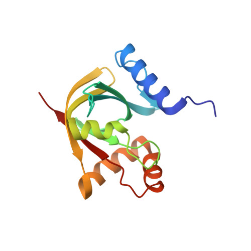
    7KL8

  • PubMed Abstract: 

    Triaza-coumarin (TA-C) is a Mycobacterium tuberculosis (Mtb) dihydrofolate reductase (DHFR) inhibitor with an IC 50 (half maximal inhibitory concentration) of ∼1 µM against the enzyme. Despite this moderate target inhibition, TA-C shows exquisite antimycobacterial activity (MIC 50 , concentration inhibiting growth by 50% = 10 to 20 nM). Here, we investigated the mechanism underlying this potency disconnect. To confirm that TA-C targets DHFR and investigate its unusual potency pattern, we focused on resistance mechanisms. In Mtb, resistance to DHFR inhibitors is frequently associated with mutations in thymidylate synthase thyA , which sensitizes Mtb to DHFR inhibition, rather than in DHFR itself. We observed thyA mutations, consistent with TA-C interfering with the folate pathway. A second resistance mechanism involved biosynthesis of the redox coenzyme F 420 Thus, we hypothesized that TA-C may be metabolized by Mtb F 420 -dependent oxidoreductases (FDORs). By chemically blocking the putative site of FDOR-mediated reduction in TA-C, we reproduced the F 420 -dependent resistance phenotype, suggesting that F 420 H 2 -dependent reduction is required for TA-C to exert its potent antibacterial activity. Indeed, chemically synthesized TA-C-Acid, the putative product of TA-C reduction, displayed a 100-fold lower IC 50 against DHFR. Screening seven recombinant Mtb FDORs revealed that at least two of these enzymes reduce TA-C. This redundancy in activation explains why no mutations in the activating enzymes were identified in the resistance screen. Analysis of the reaction products confirmed that FDORs reduce TA-C at the predicted site, yielding TA-C-Acid. This work demonstrates that intrabacterial metabolism converts TA-C, a moderately active "prodrug," into a 100-fold-more-potent DHFR inhibitor, thus explaining the disconnect between enzymatic and whole-cell activity.


  • Organizational Affiliation
    • Center for Discovery and Innovation, Hackensack Meridian Health, Nutley, NJ 07110.

Macromolecule Content 

  • Total Structure Weight: 33.64 kDa 
  • Atom Count: 2,390 
  • Modeled Residue Count: 280 
  • Deposited Residue Count: 280 
  • Unique protein chains: 1

Macromolecules

Find similar proteins by:|  3D Structure
Entity ID: 1
MoleculeChains  Sequence LengthOrganismDetailsImage
Deazaflavin-dependent nitroreductase
A, B
140Mycobacterium tuberculosisMutation(s): 0 
Gene Names: 
EC: 1 (PDB Primary Data), 1.1.98 (PDB Primary Data)
UniProt
Find proteins for P9WP11 (Mycobacterium tuberculosis (strain ATCC 25618 / H37Rv))
Explore P9WP11 
Go to UniProtKB:  P9WP11
Entity Groups
Sequence Clusters30% Identity50% Identity70% Identity90% Identity95% Identity100% Identity
UniProt GroupP9WP11
Sequence Annotations
Expand
Reference Sequence

Small Molecules

Ligands 3 Unique
IDChains Name / Formula / InChI Key2D Diagram3D Interactions
6J4

Query on 6J4


Download:Ideal Coordinates CCD File
C [auth A]COENZYME F420-3
C34 H43 N6 O21 P
YHDAXCLOUDHUAA-LROHGRLLSA-N
F42

Query on F42


Download:Ideal Coordinates CCD File
H [auth B]COENZYME F420
C29 H36 N5 O18 P
GEHSZWRGPHDXJO-NALJQGANSA-N
SO4

Query on SO4


Download:Ideal Coordinates CCD File
D [auth A]
E [auth A]
F [auth A]
G [auth A]
I [auth B]
D [auth A],
E [auth A],
F [auth A],
G [auth A],
I [auth B],
J [auth B]
SULFATE ION
O4 S
QAOWNCQODCNURD-UHFFFAOYSA-L

Experimental Data & Validation

Experimental Data

  • Method: X-RAY DIFFRACTION
  • Resolution: 2.47 Å
  • R-Value Free:  0.220 (Depositor), 0.226 (DCC) 
  • R-Value Work:  0.200 (Depositor), 0.208 (DCC) 
  • R-Value Observed: 0.201 (Depositor) 
Space Group: P 43 3 2
Unit Cell:
Length ( Å )Angle ( ˚ )
a = 169.544α = 90
b = 169.544β = 90
c = 169.544γ = 90
Software Package:
Software NamePurpose
REFMACrefinement
XDSdata reduction
Aimlessdata scaling
MOLREPphasing

Structure Validation

View Full Validation Report


Ligand Structure Quality Assessment 


Entry History 

& Funding Information

Deposition Data


Funding OrganizationLocationGrant Number
National Health and Medical Research Council (NHMRC, Australia)AustraliaAPP1128929

Revision History  (Full details and data files)

  • Version 1.0: 2021-03-24
    Type: Initial release
  • Version 1.1: 2021-10-06
    Changes: Database references
  • Version 1.2: 2023-10-18
    Changes: Data collection, Refinement description