7KDC

The complex between RhoD and the Plexin B2 RBD


Experimental Data Snapshot

  • Method: X-RAY DIFFRACTION
  • Resolution: 3.10 Å
  • R-Value Free: 0.266 
  • R-Value Work: 0.215 
  • R-Value Observed: 0.218 

wwPDB Validation   3D Report Full Report


Ligand Structure Quality Assessment 


This is version 1.3 of the entry. See complete history


Literature

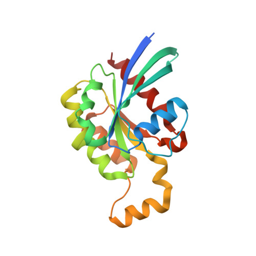
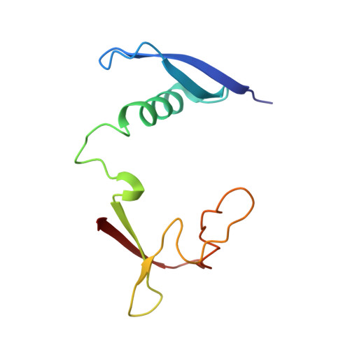
A putative structural mechanism underlying the antithetic effect of homologous RND1 and RhoD GTPases in mammalian plexin regulation.

Liu, Y.Ke, P.Kuo, Y.C.Wang, Y.Zhang, X.Song, C.Shan, Y.

(2021) Elife 10

  • DOI: https://doi.org/10.7554/eLife.64304
  • Primary Citation of Related Structures:  
    7KDC

  • PubMed Abstract: 

    Plexins are semaphorin receptors that play essential roles in mammalian neuronal axon guidance and in many other important mammalian biological processes. Plexin signaling depends on a semaphorin-induced dimerization mechanism and is modulated by small GTPases of the Rho family, of which RND1 serves as a plexin activator yet its close homolog RhoD an inhibitor. Using molecular dynamics (MD) simulations, we showed that RND1 reinforces the plexin dimerization interface, whereas RhoD destabilizes it due to their differential interaction with the cell membrane. Upon binding plexin at the Rho-GTPase-binding domain (RBD), RND1 and RhoD interact differently with the inner leaflet of the cell membrane and exert opposite effects on the dimerization interface via an allosteric network involving the RBD, RBD linkers, and a buttress segment adjacent to the dimerization interface. The differential membrane interaction is attributed to the fact that, unlike RND1, RhoD features a short C-terminal tail and a positively charged membrane interface.


  • Organizational Affiliation

    Center for Quantitative Biology, Academy for Advanced Interdisciplinary Studies, Peking University, Beijing, China.


Macromolecules
Find similar proteins by:  (by identity cutoff)  |  3D Structure
Entity ID: 1
MoleculeChains Sequence LengthOrganismDetailsImage
Rho-related GTP-binding protein RhoD
A, B
191Homo sapiensMutation(s): 2 
Gene Names: RHODARHD
UniProt & NIH Common Fund Data Resources
Find proteins for O00212 (Homo sapiens)
Explore O00212 
Go to UniProtKB:  O00212
PHAROS:  O00212
GTEx:  ENSG00000173156 
Entity Groups  
Sequence Clusters30% Identity50% Identity70% Identity90% Identity95% Identity100% Identity
UniProt GroupO00212
Sequence Annotations
Expand
  • Reference Sequence
Find similar proteins by:  (by identity cutoff)  |  3D Structure
Entity ID: 2
MoleculeChains Sequence LengthOrganismDetailsImage
Plexin-B2
C, D
103Mus musculusMutation(s): 0 
Gene Names: Plxnb2
UniProt & NIH Common Fund Data Resources
Find proteins for B2RXS4 (Mus musculus)
Explore B2RXS4 
Go to UniProtKB:  B2RXS4
IMPC:  MGI:2154239
Entity Groups  
Sequence Clusters30% Identity50% Identity70% Identity90% Identity95% Identity100% Identity
UniProt GroupB2RXS4
Sequence Annotations
Expand
  • Reference Sequence
Experimental Data & Validation

Experimental Data

  • Method: X-RAY DIFFRACTION
  • Resolution: 3.10 Å
  • R-Value Free: 0.266 
  • R-Value Work: 0.215 
  • R-Value Observed: 0.218 
  • Space Group: P 31
Unit Cell:
Length ( Å )Angle ( ˚ )
a = 82.81α = 90
b = 82.81β = 90
c = 136.761γ = 120
Software Package:
Software NamePurpose
PHENIXrefinement
HKL-2000data reduction
HKL-2000data scaling
PHASERphasing

Structure Validation

View Full Validation Report


Ligand Structure Quality Assessment 


Entry History & Funding Information

Deposition Data


Funding OrganizationLocationGrant Number
Welch FoundationUnited StatesI-1702

Revision History  (Full details and data files)

  • Version 1.0: 2021-07-28
    Type: Initial release
  • Version 1.1: 2021-08-04
    Changes: Database references
  • Version 1.2: 2021-08-11
    Changes: Database references
  • Version 1.3: 2023-10-18
    Changes: Data collection, Refinement description