


to be published
Seattle Structural Genomics Center for Infectious Disease (SSGCID), Fox III, D., Abendroth, J., DeBouver, N.D., Krysan, D.J., Lorimer, D.D., Horanyi, P.S., Edwards, T.E.To be published.
Experimental Data Snapshot
Starting Model: experimental
View more details
Entity ID: 1 | |||||
|---|---|---|---|---|---|
| Molecule | Chains | Sequence Length | Organism | Details | Image |
| Acetyl-coenzyme A synthetase | 706 | Coccidioides posadasii C735 delta SOWgp | Mutation(s): 0 Gene Names: CPC735_049370 EC: 6.2.1.1 | 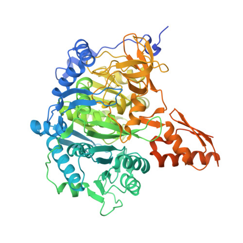 | |
UniProt | |||||
Entity Groups | |||||
| Sequence Clusters | 30% Identity50% Identity70% Identity90% Identity95% Identity100% Identity | ||||
| UniProt Group | C5PGB4 | ||||
Sequence AnnotationsExpand | |||||
Reference Sequence | |||||
| Ligands 3 Unique | |||||
|---|---|---|---|---|---|
| ID | Chains | Name / Formula / InChI Key | 2D Diagram | 3D Interactions | |
| PRX (Subject of Investigation/LOI) Download:Ideal Coordinates CCD File | B [auth A] | ADENOSINE-5'-MONOPHOSPHATE-PROPYL ESTER C13 H20 N5 O7 P XAMXMSZRQHPMRX-QYVSTXNMSA-N |  | ||
| EDO Download:Ideal Coordinates CCD File | D [auth A] E [auth A] F [auth A] G [auth A] H [auth A] | 1,2-ETHANEDIOL C2 H6 O2 LYCAIKOWRPUZTN-UHFFFAOYSA-N |  | ||
| MG Download:Ideal Coordinates CCD File | C [auth A] | MAGNESIUM ION Mg JLVVSXFLKOJNIY-UHFFFAOYSA-N |  | ||
| Length ( Å ) | Angle ( ˚ ) |
|---|---|
| a = 107.17 | α = 90 |
| b = 107.17 | β = 90 |
| c = 116.17 | γ = 120 |
| Software Name | Purpose |
|---|---|
| XSCALE | data scaling |
| PHENIX | refinement |
| PDB_EXTRACT | data extraction |
| XDS | data reduction |
| PHASER | phasing |
| Coot | model building |


