7KCB

Symmetry in Yeast Alcohol Dehydrogenase 1 -Closed Form with NAD+ and Trifluoroethanol


Experimental Data Snapshot

  • Method: ELECTRON MICROSCOPY
  • Resolution: 2.77 Å
  • Aggregation State: PARTICLE 
  • Reconstruction Method: SINGLE PARTICLE 

wwPDB Validation   3D Report Full Report


This is version 1.0 of the entry. See complete history


Literature

Cryo-Electron Microscopy Structures of Yeast Alcohol Dehydrogenase.

Guntupalli, S.R.Li, Z.Chang, L.Plapp, B.V.Subramanian, R.

(2021) Biochemistry 60: 663-677

  • DOI: https://doi.org/10.1021/acs.biochem.0c00921
  • Primary Citation of Related Structures:  
    7KC2, 7KCB, 7KCQ, 7KJY

  • PubMed Abstract: 

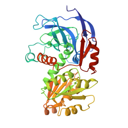
    Structures of yeast alcohol dehydrogenase determined by X-ray crystallography show that the subunits have two different conformational states in each of the two dimers that form the tetramer. Apoenzyme and holoenzyme complexes relevant to the catalytic mechanism were described, but the asymmetry led to questions about the cooperativity of the subunits in catalysis. This study used cryo-electron microscopy (cryo-EM) to provide structures for the apoenzyme, two different binary complexes with NADH, and a ternary complex with NAD + and 2,2,2-trifluoroethanol. All four subunits in each of these complexes are identical, as the tetramers have D 2 symmetry, suggesting that there is no preexisting asymmetry and that the subunits can be independently active. The apoenzyme and one enzyme-NADH complex have "open" conformations and the inverted coordination of the catalytic zinc with Cys-43, His-66, Glu-67, and Cys-153, whereas another enzyme-NADH complex and the ternary complex have closed conformations with the classical coordination of the zinc with Cys-43, His-66, Cys-153, and a water or the oxygen of trifluoroethanol. The conformational change involves interactions of Arg-340 with the pyrophosphate group of the coenzyme and Glu-67. The cryo-EM and X-ray crystallography studies provide structures relevant for the catalytic mechanism.


  • Organizational Affiliation

    Institute for Stem Cell Science and Regenerative Medicine, Bangalore, India.


Macromolecules
Find similar proteins by:  (by identity cutoff)  |  3D Structure
Entity ID: 1
MoleculeChains Sequence LengthOrganismDetailsImage
ADH1 isoform 1
A, B, C, D
347Saccharomyces cerevisiaeMutation(s): 0 
UniProt
Find proteins for P00330 (Saccharomyces cerevisiae (strain ATCC 204508 / S288c))
Explore P00330 
Go to UniProtKB:  P00330
Entity Groups  
Sequence Clusters30% Identity50% Identity70% Identity90% Identity95% Identity100% Identity
UniProt GroupP00330
Sequence Annotations
Expand
  • Reference Sequence
Small Molecules
Ligands 3 Unique
IDChains Name / Formula / InChI Key2D Diagram3D Interactions
NAD (Subject of Investigation/LOI)
Query on NAD

Download Ideal Coordinates CCD File 
G [auth A],
K [auth B],
O [auth C],
S [auth D]
NICOTINAMIDE-ADENINE-DINUCLEOTIDE
C21 H27 N7 O14 P2
BAWFJGJZGIEFAR-NNYOXOHSSA-N
ETF (Subject of Investigation/LOI)
Query on ETF

Download Ideal Coordinates CCD File 
H [auth A],
L [auth B],
P [auth C],
T [auth D]
TRIFLUOROETHANOL
C2 H3 F3 O
RHQDFWAXVIIEBN-UHFFFAOYSA-N
ZN (Subject of Investigation/LOI)
Query on ZN

Download Ideal Coordinates CCD File 
E [auth A]
F [auth A]
I [auth B]
J [auth B]
M [auth C]
E [auth A],
F [auth A],
I [auth B],
J [auth B],
M [auth C],
N [auth C],
Q [auth D],
R [auth D]
ZINC ION
Zn
PTFCDOFLOPIGGS-UHFFFAOYSA-N
Experimental Data & Validation

Experimental Data

  • Method: ELECTRON MICROSCOPY
  • Resolution: 2.77 Å
  • Aggregation State: PARTICLE 
  • Reconstruction Method: SINGLE PARTICLE 
EM Software:
TaskSoftware PackageVersion
MODEL REFINEMENTPHENIX1.17
RECONSTRUCTIONcryoSPARC2

Structure Validation

View Full Validation Report


Entry History 

Deposition Data

Revision History  (Full details and data files)

  • Version 1.0: 2021-03-31
    Type: Initial release