7K96 | pdb_00007k96

Human DNA polymerase beta ternary complex with templating cytosine and incoming deoxyguanosine diphosphate


Experimental Data Snapshot

  • Method: X-RAY DIFFRACTION
  • Resolution: 2.10 Å
  • R-Value Free: 
    0.246 (Depositor), 0.247 (DCC) 
  • R-Value Work: 
    0.183 (Depositor), 0.184 (DCC) 
  • R-Value Observed: 
    0.189 (Depositor) 

Starting Model: experimental
View more details

wwPDB Validation 3D Report Full Report

Validation slider image for 7K96

Ligand Structure Quality Assessment 


This is version 1.2 of the entry. See complete history

Literature

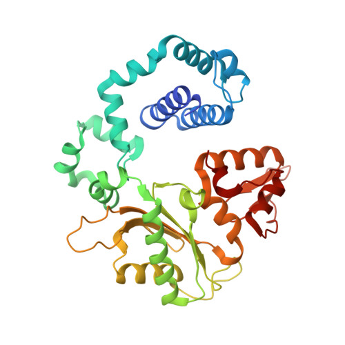
Mechanism of Deoxyguanosine Diphosphate Insertion by Human DNA Polymerase beta.

Varela, F.A.Freudenthal, B.D.

(2021) Biochemistry 60: 373-380

  • DOI: https://doi.org/10.1021/acs.biochem.0c00847
  • Primary Citation Related Structures: 
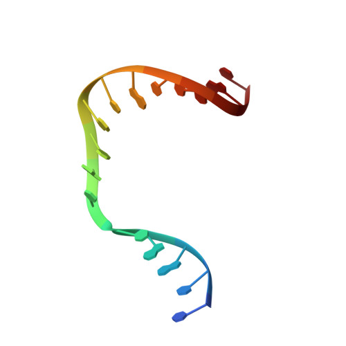
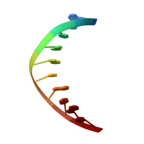
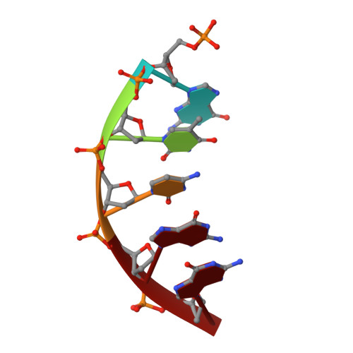
    7K96, 7K97

  • PubMed Abstract: 

    DNA polymerases play vital roles in the maintenance and replication of genomic DNA by synthesizing new nucleotide polymers using nucleoside triphosphates as substrates. Deoxynucleoside triphosphates (dNTPs) are the canonical substrates for DNA polymerases; however, some bacterial polymerases have been demonstrated to insert deoxynucleoside diphosphates (dNDPs), which lack a third phosphate group, the γ-phosphate. Whether eukaryotic polymerases can efficiently incorporate dNDPs has not been investigated, and much about the chemical or structural role played by the γ-phosphate of dNTPs remains unknown. Using the model mammalian polymerase (Pol) β, we examine how Pol β incorporates a substrate lacking a γ-phosphate [deoxyguanosine diphosphate (dGDP)] utilizing kinetic and crystallographic approaches. Using single-turnover kinetics, we determined dGDP insertion across a templating dC by Pol β to be drastically impaired when compared to dGTP insertion. We found the most significant impairment in the apparent insertion rate ( k pol ), which was reduced 32000-fold compared to that of dGTP insertion. X-ray crystal structures revealed similar enzyme-substrate contacts for both dGDP and dGTP. These findings suggest the insertion efficiency of dGDP is greatly decreased due to impairments in polymerase chemistry. This work is the first instance of a mammalian polymerase inserting a diphosphate nucleotide and provides insight into the nature of polymerase mechanisms by highlighting how these enzymes have evolved to use triphosphate nucleotide substrates.


  • Organizational Affiliation
    • Department of Biochemistry and Molecular Biology and Department of Cancer Biology, University of Kansas Medical Center, Kansas City, Kansas 66160, United States.

Macromolecule Content 

  • Total Structure Weight: 48.87 kDa 
  • Atom Count: 3,491 
  • Modeled Residue Count: 357 
  • Deposited Residue Count: 366 
  • Unique protein chains: 1
  • Unique nucleic acid chains: 3

Macromolecules


Find similar proteins by:|  3D Structure
Entity ID: 4
MoleculeChains  Sequence LengthOrganismDetailsImage
DNA polymerase betaD [auth A]335Homo sapiensMutation(s): 0 
Gene Names: POLB
EC: 2.7.7.7 (PDB Primary Data), 4.2.99 (PDB Primary Data), 4.2.99.18 (UniProt)
UniProt & NIH Common Fund Data Resources
Find proteins for P06746 (Homo sapiens)
Explore P06746 
Go to UniProtKB:  P06746
PHAROS:  P06746
GTEx:  ENSG00000070501 
Entity Groups
Sequence Clusters30% Identity50% Identity70% Identity90% Identity95% Identity100% Identity
UniProt GroupP06746
Sequence Annotations
Expand
Reference Sequence
Find similar nucleic acids by:  Sequence
Entity ID: 1
MoleculeChains LengthOrganismImage
DNA (5'-D(*CP*CP*GP*AP*CP*CP*GP*CP*GP*CP*AP*TP*CP*AP*GP*C)-3')A [auth T]16synthetic construct
Sequence Annotations
Expand
Reference Sequence
Find similar nucleic acids by:  Sequence
Entity ID: 2
MoleculeChains LengthOrganismImage
DNA (5'-D(*GP*CP*TP*GP*AP*TP*GP*CP*GP*C)-3')B [auth P]10synthetic construct
Sequence Annotations
Expand
Reference Sequence
Find similar nucleic acids by:  Sequence
Entity ID: 3
MoleculeChains LengthOrganismImage
DNA (5'-D(P*GP*TP*CP*GP*G)-3')C [auth D]5synthetic construct
Sequence Annotations
Expand
Reference Sequence

Small Molecules

Ligands 5 Unique
IDChains Name / Formula / InChI Key2D Diagram3D Interactions
DGI
(Subject of Investigation/LOI)

Query on DGI


Download:Ideal Coordinates CCD File
V [auth A]2'-DEOXYGUANOSINE-5'-DIPHOSPHATE
C10 H15 N5 O10 P2
CIKGWCTVFSRMJU-KVQBGUIXSA-N
EDO

Query on EDO


Download:Ideal Coordinates CCD File
G [auth A],
W [auth A],
X [auth A],
Y [auth A]
1,2-ETHANEDIOL
C2 H6 O2
LYCAIKOWRPUZTN-UHFFFAOYSA-N
CA

Query on CA


Download:Ideal Coordinates CCD File
H [auth A],
I [auth A],
J [auth A]
CALCIUM ION
Ca
BHPQYMZQTOCNFJ-UHFFFAOYSA-N
CL

Query on CL


Download:Ideal Coordinates CCD File
E [auth T]
N [auth A]
O [auth A]
P [auth A]
Q [auth A]
E [auth T],
N [auth A],
O [auth A],
P [auth A],
Q [auth A],
R [auth A],
S [auth A],
T [auth A],
U [auth A]
CHLORIDE ION
Cl
VEXZGXHMUGYJMC-UHFFFAOYSA-M
NA

Query on NA


Download:Ideal Coordinates CCD File
F [auth P],
K [auth A],
L [auth A],
M [auth A]
SODIUM ION
Na
FKNQFGJONOIPTF-UHFFFAOYSA-N

Experimental Data & Validation

Experimental Data

  • Method: X-RAY DIFFRACTION
  • Resolution: 2.10 Å
  • R-Value Free:  0.246 (Depositor), 0.247 (DCC) 
  • R-Value Work:  0.183 (Depositor), 0.184 (DCC) 
  • R-Value Observed: 0.189 (Depositor) 
Space Group: P 1 21 1
Unit Cell:
Length ( Å )Angle ( ˚ )
a = 50.8α = 90
b = 80.4β = 107.9
c = 55.5γ = 90
Software Package:
Software NamePurpose
PHENIXrefinement
HKL-3000data scaling
HKL-3000data reduction
PHASERphasing

Structure Validation

View Full Validation Report


Ligand Structure Quality Assessment 


Entry History 

& Funding Information

Deposition Data


Funding OrganizationLocationGrant Number
National Institutes of Health/National Institute of General Medical Sciences (NIH/NIGMS)United States--

Revision History  (Full details and data files)

  • Version 1.0: 2021-02-03
    Type: Initial release
  • Version 1.1: 2021-02-24
    Changes: Database references
  • Version 1.2: 2023-10-18
    Changes: Data collection, Database references, Refinement description