7K3G | pdb_00007k3g

SARS-CoV-2 Envelope Protein Transmembrane Domain: Pentameric Structure Determined by Solid-State NMR


Experimental Data Snapshot

  • Method: SOLID-STATE NMR
  • Conformers Calculated: 192 
  • Conformers Submitted: 10 
  • Selection Criteria: structures with the lowest energy 

wwPDB Validation 3D Report Full Report

Validation slider image for 7K3G

This is version 2.5 of the entry. See complete history

Literature

Structure and drug binding of the SARS-CoV-2 envelope protein transmembrane domain in lipid bilayers.

Mandala, V.S.McKay, M.J.Shcherbakov, A.A.Dregni, A.J.Kolocouris, A.Hong, M.

(2020) Nat Struct Mol Biol 27: 1202-1208

  • DOI: https://doi.org/10.1038/s41594-020-00536-8
  • Primary Citation Related Structures: 
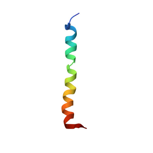
    7K3G

  • PubMed Abstract: 

    An essential protein of the SARS-CoV-2 virus, the envelope protein E, forms a homopentameric cation channel that is important for virus pathogenicity. Here we report a 2.1-Å structure and the drug-binding site of E's transmembrane domain (ETM), determined using solid-state NMR spectroscopy. In lipid bilayers that mimic the endoplasmic reticulum-Golgi intermediate compartment (ERGIC) membrane, ETM forms a five-helix bundle surrounding a narrow pore. The protein deviates from the ideal α-helical geometry due to three phenylalanine residues, which stack within each helix and between helices. Together with valine and leucine interdigitation, these cause a dehydrated pore compared with the viroporins of influenza viruses and HIV. Hexamethylene amiloride binds the polar amino-terminal lumen, whereas acidic pH affects the carboxy-terminal conformation. Thus, the N- and C-terminal halves of this bipartite channel may interact with other viral and host proteins semi-independently. The structure sets the stage for designing E inhibitors as antiviral drugs.


  • Organizational Affiliation
    • Department of Chemistry, Massachusetts Institute of Technology, Cambridge, MA, USA.

Macromolecule Content 

  • Total Structure Weight: 16.81 kDa 
  • Atom Count: 1,190 
  • Modeled Residue Count: 155 
  • Deposited Residue Count: 155 
  • Unique protein chains: 1

Macromolecules

Find similar proteins by:|  3D Structure
Entity ID: 1
MoleculeChains  Sequence LengthOrganismDetailsImage
Envelope small membrane protein
A, B, C, D, E
31Severe acute respiratory syndrome coronavirus 2Mutation(s): 0 
Gene Names: E4
Membrane Entity: Yes 
UniProt
Find proteins for P0DTC4 (Severe acute respiratory syndrome coronavirus 2)
Explore P0DTC4 
Go to UniProtKB:  P0DTC4
Entity Groups
Sequence Clusters30% Identity50% Identity70% Identity90% Identity95% Identity100% Identity
UniProt GroupP0DTC4
Sequence Annotations
Expand
Reference Sequence

Experimental Data & Validation

Experimental Data

  • Method: SOLID-STATE NMR
  • Conformers Calculated: 192 
  • Conformers Submitted: 10 
  • Selection Criteria: structures with the lowest energy 

Structure Validation

View Full Validation Report


Entry History 

& Funding Information

Deposition Data


Funding OrganizationLocationGrant Number
National Institutes of Health/National Institute of General Medical Sciences (NIH/NIGMS)United StatesGM088204

Revision History  (Full details and data files)

  • Version 1.0: 2020-09-30
    Type: Initial release
  • Version 2.0: 2020-10-21
    Type: Coordinate replacement
    Reason: Polymer geometry
    Changes: Atomic model, Data collection, Derived calculations
  • Version 2.1: 2020-10-28
    Changes: Database references
  • Version 2.2: 2020-11-25
    Changes: Database references
  • Version 2.3: 2020-12-16
    Changes: Database references
  • Version 2.4: 2023-06-14
    Changes: Database references, Other
  • Version 2.5: 2024-05-15
    Changes: Data collection, Database references