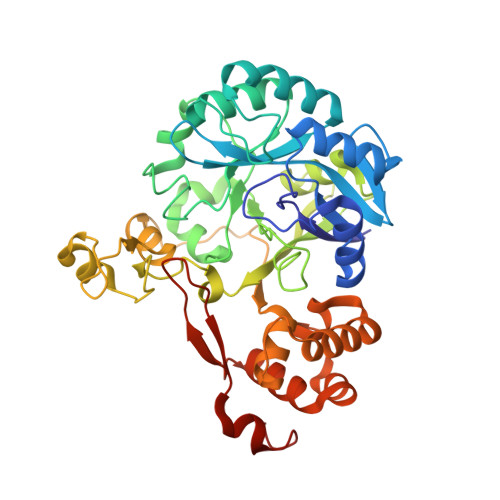
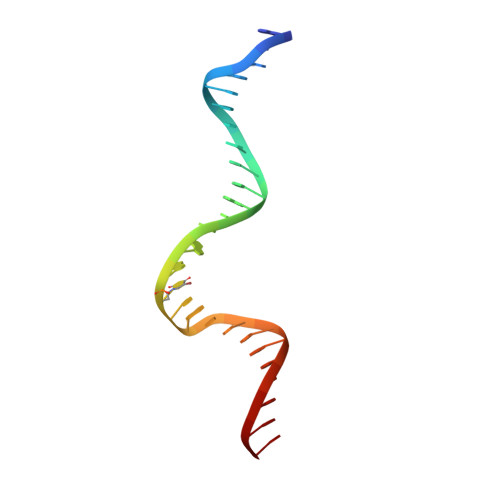
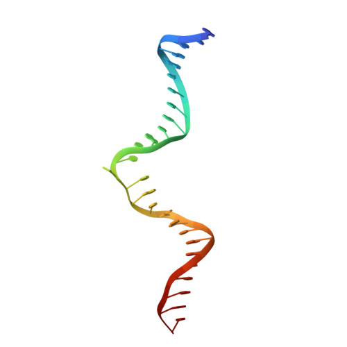
7K30

Crystal structure of Endonuclease Q complex with 27-mer duplex substrate with dU at the active site


Experimental Data Snapshot

  • Method: X-RAY DIFFRACTION
  • Resolution: 2.34 Å
  • R-Value Free: 0.228 
  • R-Value Work: 0.205 
  • R-Value Observed: 0.207 

Starting Model: experimental
View more details

wwPDB Validation   3D Report Full Report


This is version 1.1 of the entry. See complete history


Literature

Structural basis for recognition of distinct deaminated DNA lesions by endonuclease Q.

Shi, K.Moeller, N.H.Banerjee, S.McCann, J.L.Carpenter, M.A.Yin, L.Moorthy, R.Orellana, K.Harki, D.A.Harris, R.S.Aihara, H.

(2021) Proc Natl Acad Sci U S A 118

  • DOI: https://doi.org/10.1073/pnas.2021120118
  • Primary Citation of Related Structures:  
    7K30, 7K31, 7K32, 7K33

  • PubMed Abstract: 

    Spontaneous deamination of DNA cytosine and adenine into uracil and hypoxanthine, respectively, causes C to T and A to G transition mutations if left unrepaired. Endonuclease Q (EndoQ) initiates the repair of these premutagenic DNA lesions in prokaryotes by cleaving the phosphodiester backbone 5' of either uracil or hypoxanthine bases or an apurinic/apyrimidinic (AP) lesion generated by the excision of these damaged bases. To understand how EndoQ achieves selectivity toward these structurally diverse substrates without cleaving undamaged DNA, we determined the crystal structures of Pyrococcus furiosus EndoQ bound to DNA substrates containing uracil, hypoxanthine, or an AP lesion. The structures show that substrate engagement by EndoQ depends both on a highly distorted conformation of the DNA backbone, in which the target nucleotide is extruded out of the helix, and direct hydrogen bonds with the deaminated bases. A concerted swing motion of the zinc-binding and C-terminal helical domains of EndoQ toward its catalytic domain allows the enzyme to clamp down on a sharply bent DNA substrate, shaping a deep active-site pocket that accommodates the extruded deaminated base. Within this pocket, uracil and hypoxanthine bases interact with distinct sets of amino acid residues, with positioning mediated by an essential magnesium ion. The EndoQ-DNA complex structures reveal a unique mode of damaged DNA recognition and provide mechanistic insights into the initial step of DNA damage repair by the alternative excision repair pathway. Furthermore, we demonstrate that the unique activity of EndoQ is useful for studying DNA deamination and repair in mammalian systems.


  • Organizational Affiliation

    Department of Biochemistry, Molecular Biology, and Biophysics, University of Minnesota, Minneapolis, MN 55455.


Macromolecules

Find similar proteins by:  (by identity cutoff)  |  3D Structure
Entity ID: 1
MoleculeChains Sequence LengthOrganismDetailsImage
Endonuclease Q400Pyrococcus furiosusMutation(s): 1 
UniProt
Find proteins for Q8U0N5 (Pyrococcus furiosus (strain ATCC 43587 / DSM 3638 / JCM 8422 / Vc1))
Explore Q8U0N5 
Go to UniProtKB:  Q8U0N5
Entity Groups  
Sequence Clusters30% Identity50% Identity70% Identity90% Identity95% Identity100% Identity
UniProt GroupQ8U0N5
Sequence Annotations
Expand
  • Reference Sequence
Find similar nucleic acids by:  (by identity cutoff)  |  3D Structure
Entity ID: 2
MoleculeChains LengthOrganismImage
DNA (27-MER)27synthetic construct
Sequence Annotations
Expand
  • Reference Sequence
Find similar nucleic acids by:  (by identity cutoff)  |  3D Structure
Entity ID: 3
MoleculeChains LengthOrganismImage
DNA (27-MER)27synthetic construct
Sequence Annotations
Expand
  • Reference Sequence
Small Molecules
Ligands 3 Unique
IDChains Name / Formula / InChI Key2D Diagram3D Interactions
ZN
Query on ZN

Download Ideal Coordinates CCD File 
D [auth A],
E [auth A]
ZINC ION
Zn
PTFCDOFLOPIGGS-UHFFFAOYSA-N
EDO
Query on EDO

Download Ideal Coordinates CCD File 
G [auth A],
H [auth A],
I [auth A],
J [auth A]
1,2-ETHANEDIOL
C2 H6 O2
LYCAIKOWRPUZTN-UHFFFAOYSA-N
MG
Query on MG

Download Ideal Coordinates CCD File 
F [auth A]MAGNESIUM ION
Mg
JLVVSXFLKOJNIY-UHFFFAOYSA-N
Experimental Data & Validation

Experimental Data

  • Method: X-RAY DIFFRACTION
  • Resolution: 2.34 Å
  • R-Value Free: 0.228 
  • R-Value Work: 0.205 
  • R-Value Observed: 0.207 
  • Space Group: H 3
Unit Cell:
Length ( Å )Angle ( ˚ )
a = 152.7α = 90
b = 152.7β = 90
c = 118.17γ = 120
Software Package:
Software NamePurpose
PHENIXrefinement
XDSdata reduction
Aimlessdata scaling
PHASERphasing

Structure Validation

View Full Validation Report


Entry History & Funding Information

Deposition Data


Funding OrganizationLocationGrant Number
National Institutes of Health/National Institute of General Medical Sciences (NIH/NIGMS)United StatesGM118047

Revision History  (Full details and data files)

  • Version 1.0: 2021-03-17
    Type: Initial release
  • Version 1.1: 2023-10-18
    Changes: Data collection, Database references, Refinement description