7K0R | pdb_00007k0r

Nucleotide bound SARS-CoV-2 Nsp15


Experimental Data Snapshot

  • Method: ELECTRON MICROSCOPY
  • Resolution: 3.30 Å
  • Aggregation State: PARTICLE 
  • Reconstruction Method: SINGLE PARTICLE 

wwPDB Validation 3D Report Full Report

Validation slider image for 7K0R

This is version 1.3 of the entry. See complete history

Literature

Cryo-EM structures of the SARS-CoV-2 endoribonuclease Nsp15 reveal insight into nuclease specificity and dynamics.

Pillon, M.C.Frazier, M.N.Dillard, L.B.Williams, J.G.Kocaman, S.Krahn, J.M.Perera, L.Hayne, C.K.Gordon, J.Stewart, Z.D.Sobhany, M.Deterding, L.J.Hsu, A.L.Dandey, V.P.Borgnia, M.J.Stanley, R.E.

(2021) Nat Commun 12: 636-636

  • DOI: https://doi.org/10.1038/s41467-020-20608-z
  • Primary Citation Related Structures: 
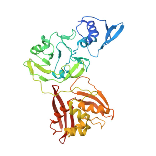
    7K0R

  • PubMed Abstract: 

    Nsp15, a uridine specific endoribonuclease conserved across coronaviruses, processes viral RNA to evade detection by host defense systems. Crystal structures of Nsp15 from different coronaviruses have shown a common hexameric assembly, yet how the enzyme recognizes and processes RNA remains poorly understood. Here we report a series of cryo-EM reconstructions of SARS-CoV-2 Nsp15, in both apo and UTP-bound states. The cryo-EM reconstructions, combined with biochemistry, mass spectrometry, and molecular dynamics, expose molecular details of how critical active site residues recognize uridine and facilitate catalysis of the phosphodiester bond. Mass spectrometry revealed the accumulation of cyclic phosphate cleavage products, while analysis of the apo and UTP-bound datasets revealed conformational dynamics not observed by crystal structures that are likely important to facilitate substrate recognition and regulate nuclease activity. Collectively, these findings advance understanding of how Nsp15 processes viral RNA and provide a structural framework for the development of new therapeutics.


  • Organizational Affiliation
    • Signal Transduction Laboratory, National Institute of Environmental Health Sciences, National Institutes of Health, Department of Health and Human Services, 111 T. W. Alexander Drive, Research Triangle Park, NC, 27709, USA. monica.pillon@nih.gov.

Macromolecule Content 

  • Total Structure Weight: 247.1 kDa 
  • Atom Count: 16,038 
  • Modeled Residue Count: 2,070 
  • Deposited Residue Count: 2,172 
  • Unique protein chains: 1

Macromolecules

Find similar proteins by:|  3D Structure
Entity ID: 1
MoleculeChains  Sequence LengthOrganismDetailsImage
Uridylate-specific endoribonuclease
A, B, C, D, E
A, B, C, D, E, F
362Severe acute respiratory syndrome coronavirus 2Mutation(s): 0 
Gene Names: rep1a-1b
EC: 3.1
UniProt
Find proteins for P0DTD1 (Severe acute respiratory syndrome coronavirus 2)
Explore P0DTD1 
Go to UniProtKB:  P0DTD1
Entity Groups
Sequence Clusters30% Identity50% Identity70% Identity90% Identity95% Identity100% Identity
UniProt GroupP0DTD1
Sequence Annotations
Expand
Reference Sequence

Small Molecules

Ligands 2 Unique
IDChains Name / Formula / InChI Key2D Diagram3D Interactions
U5P
(Subject of Investigation/LOI)

Query on U5P


Download:Ideal Coordinates CCD File
G [auth A]
I [auth B]
K [auth C]
M [auth D]
O [auth E]
G [auth A],
I [auth B],
K [auth C],
M [auth D],
O [auth E],
Q [auth F]
URIDINE-5'-MONOPHOSPHATE
C9 H13 N2 O9 P
DJJCXFVJDGTHFX-XVFCMESISA-N
PO4

Query on PO4


Download:Ideal Coordinates CCD File
H [auth A]
J [auth B]
L [auth C]
N [auth D]
P [auth E]
H [auth A],
J [auth B],
L [auth C],
N [auth D],
P [auth E],
R [auth F]
PHOSPHATE ION
O4 P
NBIIXXVUZAFLBC-UHFFFAOYSA-K

Experimental Data & Validation

Experimental Data

  • Method: ELECTRON MICROSCOPY
  • Resolution: 3.30 Å
  • Aggregation State: PARTICLE 
  • Reconstruction Method: SINGLE PARTICLE 

Structure Validation

View Full Validation Report


Entry History 

& Funding Information

Deposition Data


Funding OrganizationLocationGrant Number
National Institutes of Health/National Institute of Environmental Health Sciences (NIH/NIEHS)United States--

Revision History  (Full details and data files)

  • Version 1.0: 2020-12-09
    Type: Initial release
  • Version 1.1: 2021-01-20
    Changes: Database references, Source and taxonomy, Structure summary
  • Version 1.2: 2021-02-17
    Changes: Database references
  • Version 1.3: 2024-03-06
    Changes: Data collection, Database references