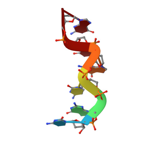
Water structure around a left-handed Z-DNA fragment analyzed by cryo neutron crystallography.
Harp, J.M., Coates, L., Sullivan, B., Egli, M.(2021) Nucleic Acids Res 49: 4782-4792
- PubMed: 33872377 Search on PubMedSearch on PubMed Central
- DOI: https://doi.org/10.1093/nar/gkab264
- Primary Citation Related Structures:
7JY2 - PubMed Abstract:
Even in high-quality X-ray crystal structures of oligonucleotides determined at a resolution of 1 Å or higher, the orientations of first-shell water molecules remain unclear. We used cryo neutron crystallography to gain insight into the H-bonding patterns of water molecules around the left-handed Z-DNA duplex [d(CGCGCG)]2. The neutron density visualized at 1.5 Å resolution for the first time allows us to pinpoint the orientations of most of the water molecules directly contacting the DNA and of many second-shell waters. In particular, H-bond acceptor and donor patterns for water participating in prominent hydration motifs inside the minor groove, on the convex surface or bridging nucleobase and phosphate oxygen atoms are finally revealed. Several water molecules display entirely unexpected orientations. For example, a water molecule located at H-bonding distance from O6 keto oxygen atoms of two adjacent guanines directs both its deuterium atoms away from the keto groups. Exocyclic amino groups of guanine (N2) and cytosine (N4) unexpectedly stabilize waters H-bonded to O2 keto oxygens from adjacent cytosines and O6 keto oxygens from adjacent guanines, respectively. Our structure offers the most detailed view to date of DNA solvation in the solid-state undistorted by metal ions or polyamines.
- Department of Biochemistry and Center for Structural Biology, Vanderbilt University, School of Medicine, Nashville, TN 37232, USA.
Organizational Affiliation: