7JX9

The crystal structure of human ornithine aminotransferase with an intermediate bound during inactivation by (1S,3S)-3-amino-4-(hexafluoropropan-2-ylidenyl)-cyclopentane-1-carboxylic acid.


Experimental Data Snapshot

  • Method: X-RAY DIFFRACTION
  • Resolution: 1.96 Å
  • R-Value Free: 0.213 
  • R-Value Work: 0.186 
  • R-Value Observed: 0.187 

wwPDB Validation   3D Report Full Report


Ligand Structure Quality Assessment 


This is version 1.2 of the entry. See complete history


Literature

Structural and Kinetic Analyses Reveal the Dual Inhibition Modes of Ornithine Aminotransferase by (1 S ,3 S )-3-Amino-4-(hexafluoropropan-2-ylidenyl)-cyclopentane-1-carboxylic Acid (BCF 3 ).

Butrin, A.Beaupre, B.A.Kadamandla, N.Zhao, P.Shen, S.Silverman, R.B.Moran, G.R.Liu, D.

(2021) ACS Chem Biol 16: 67-75

  • DOI: https://doi.org/10.1021/acschembio.0c00728
  • Primary Citation of Related Structures:  
    7JX9

  • PubMed Abstract: 

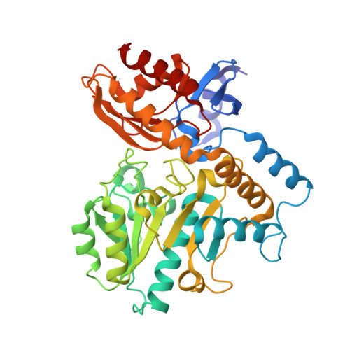
    Hepatocellular carcinoma (HCC) is the most common form of liver cancer and the leading cause of death among people with cirrhosis. HCC is typically diagnosed in advanced stages when tumors are resistant to both radio- and chemotherapy. Human ornithine aminotransferase ( h OAT) is a pyridoxal-5'-phosphate (PLP)-dependent enzyme involved in glutamine and proline metabolism. Because h OAT is overexpressed in HCC cells and a contributing factor for the uncontrolled cellular division that propagates malignant tumors (Ueno et al. J. Hepatol. 2014, 61, 1080-1087), it is a potential drug target for the treatment of HCC. (1 S ,3 S )-3-Amino-4-(hexafluoropropan-2-ylidenyl)-cyclopentane-1-carboxylic acid (BCF 3 ) has been shown in animal models to slow the progression of HCC by acting as a selective and potent mechanism-based inactivator of OAT (Zigmond et al. ACS Med. Chem. Lett. 2015, 6, 840-844). Previous studies have shown that the BCF 3 - h OAT reaction has a bifurcation in which only 8% of the inhibitor inactivates the enzyme while the remaining 92% ultimately acts as a substrate and undergoes hydrolysis to regenerate the active PLP form of the enzyme. In this manuscript, the rate-limiting step of the inactivation mechanism was determined by stopped-flow spectrophotometry and time-dependent 19 F NMR experiments to be the decay of a long-lived external aldimine species. A crystal structure of this transient complex revealed both the structural basis for fractional irreversible inhibition and the principal mode of inhibition of h OAT by BCF 3 , which is to trap the enzyme in this transient but quasi-stable external aldimine form.


  • Organizational Affiliation

    Department of Chemistry and Biochemistry, Loyola University Chicago, 1068 W Sheridan Rd, Chicago, Illinois 60660, United States.


Macromolecules
Find similar proteins by:  (by identity cutoff)  |  3D Structure
Entity ID: 1
MoleculeChains Sequence LengthOrganismDetailsImage
Ornithine aminotransferase, mitochondrial
A, B, C
404Homo sapiensMutation(s): 0 
Gene Names: OAT
EC: 2.6.1.13
UniProt & NIH Common Fund Data Resources
Find proteins for P04181 (Homo sapiens)
Explore P04181 
Go to UniProtKB:  P04181
PHAROS:  P04181
GTEx:  ENSG00000065154 
Entity Groups  
Sequence Clusters30% Identity50% Identity70% Identity90% Identity95% Identity100% Identity
UniProt GroupP04181
Sequence Annotations
Expand
  • Reference Sequence
Small Molecules
Ligands 2 Unique
IDChains Name / Formula / InChI Key2D Diagram3D Interactions
IF1 (Subject of Investigation/LOI)
Query on IF1

Download Ideal Coordinates CCD File 
D [auth A],
F [auth B],
G [auth C]
(1S,3S,4S)-3-[(E)-({3-hydroxy-2-methyl-5-[(phosphonooxy)methyl]pyridin-4-yl}methylidene)amino]-4-(1,1,3,3,3-pentafluoroprop-1-en-2-yl)cyclopentane-1-carboxylic acid
C17 H18 F5 N2 O7 P
NXIZEOJYEPCEAL-UJKLFKHKSA-N
VLS
Query on VLS

Download Ideal Coordinates CCD File 
E [auth A],
H [auth C]
N-[1,3-dihydroxy-2-(hydroxymethyl)propan-2-yl]glycine
C6 H13 N O5
SEQKRHFRPICQDD-UHFFFAOYSA-N
Experimental Data & Validation

Experimental Data

  • Method: X-RAY DIFFRACTION
  • Resolution: 1.96 Å
  • R-Value Free: 0.213 
  • R-Value Work: 0.186 
  • R-Value Observed: 0.187 
  • Space Group: P 32 2 1
Unit Cell:
Length ( Å )Angle ( ˚ )
a = 115.304α = 90
b = 115.304β = 90
c = 185.431γ = 120
Software Package:
Software NamePurpose
XDSdata reduction
XSCALEdata scaling
PHENIXrefinement
PDB_EXTRACTdata extraction
PHASERphasing

Structure Validation

View Full Validation Report


Ligand Structure Quality Assessment 


Entry History & Funding Information

Deposition Data


Funding OrganizationLocationGrant Number
National Institutes of Health/National Institute on Drug Abuse (NIH/NIDA)United StatesR01 DA030604

Revision History  (Full details and data files)

  • Version 1.0: 2021-01-06
    Type: Initial release
  • Version 1.1: 2021-01-27
    Changes: Database references
  • Version 1.2: 2023-10-18
    Changes: Data collection, Database references, Refinement description