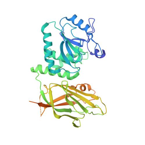
7JVX

Crystal structure of PTEN (aa 7-353 followed by spacer TGGGSGGTGGGSGGTGGGCY ligated to peptide pSDpTpTDpSDPENEPFDED)


Experimental Data Snapshot

  • Method: X-RAY DIFFRACTION
  • Resolution: 3.20 Å
  • R-Value Free: 0.296 
  • R-Value Work: 0.244 
  • R-Value Observed: 0.247 

Starting Model: experimental
View more details

wwPDB Validation   3D Report Full Report


This is version 1.2 of the entry. See complete history


Literature

The structural basis of PTEN regulation by multi-site phosphorylation.

Dempsey, D.R.Viennet, T.Iwase, R.Park, E.Henriquez, S.Chen, Z.Jeliazkov, J.R.Palanski, B.A.Phan, K.L.Coote, P.Gray, J.J.Eck, M.J.Gabelli, S.B.Arthanari, H.Cole, P.A.

(2021) Nat Struct Mol Biol 28: 858-868

  • DOI: https://doi.org/10.1038/s41594-021-00668-5
  • Primary Citation of Related Structures:  
    7JTX, 7JUK, 7JUL, 7JVX

  • PubMed Abstract: 

    Phosphatase and tensin homolog (PTEN) is a phosphatidylinositol-3,4,5-triphosphate (PIP 3 ) phospholipid phosphatase that is commonly mutated or silenced in cancer. PTEN's catalytic activity, cellular membrane localization and stability are orchestrated by a cluster of C-terminal phosphorylation (phospho-C-tail) events on Ser380, Thr382, Thr383 and Ser385, but the molecular details of this multi-faceted regulation have remained uncertain. Here we use a combination of protein semisynthesis, biochemical analysis, NMR, X-ray crystallography and computational simulations on human PTEN and its sea squirt homolog, VSP, to obtain a detailed picture of how the phospho-C-tail forms a belt around the C2 and phosphatase domains of PTEN. We also visualize a previously proposed dynamic N-terminal α-helix and show that it is key for PTEN catalysis but disordered upon phospho-C-tail interaction. This structural model provides a comprehensive framework for how C-tail phosphorylation can impact PTEN's cellular functions.


  • Organizational Affiliation

    Division of Genetics, Department of Medicine, Brigham and Women's Hospital, Boston, MA, USA.


Macromolecules
Find similar proteins by:  (by identity cutoff)  |  3D Structure
Entity ID: 1
MoleculeChains Sequence LengthOrganismDetailsImage
Phosphatidylinositol 3,4,5-trisphosphate 3-phosphatase and dual-specificity protein phosphatase PTEN422Homo sapiensMutation(s): 0 
Gene Names: PTENMMAC1TEP1
EC: 3.1.3.16 (PDB Primary Data), 3.1.3.48 (PDB Primary Data), 3.1.3.67 (PDB Primary Data), 3.1.3 (UniProt)
UniProt & NIH Common Fund Data Resources
Find proteins for P60484 (Homo sapiens)
Explore P60484 
Go to UniProtKB:  P60484
PHAROS:  P60484
GTEx:  ENSG00000171862 
Entity Groups  
Sequence Clusters30% Identity50% Identity70% Identity90% Identity95% Identity100% Identity
UniProt GroupP60484
Sequence Annotations
Expand
  • Reference Sequence
Experimental Data & Validation

Experimental Data

  • Method: X-RAY DIFFRACTION
  • Resolution: 3.20 Å
  • R-Value Free: 0.296 
  • R-Value Work: 0.244 
  • R-Value Observed: 0.247 
  • Space Group: I 4
Unit Cell:
Length ( Å )Angle ( ˚ )
a = 113.39α = 90
b = 113.39β = 90
c = 57.9γ = 90
Software Package:
Software NamePurpose
REFMACrefinement
XDSdata reduction
Aimlessdata scaling
PDB_EXTRACTdata extraction
REFMACphasing

Structure Validation

View Full Validation Report


Entry History & Funding Information

Deposition Data


Funding OrganizationLocationGrant Number
National Institutes of Health/National Cancer Institute (NIH/NCI)United States--
National Institutes of Health/National Institute of General Medical Sciences (NIH/NIGMS)United StatesK99GM130961

Revision History  (Full details and data files)

  • Version 1.0: 2021-08-04
    Type: Initial release
  • Version 1.1: 2021-10-20
    Changes: Database references
  • Version 1.2: 2023-10-18
    Changes: Data collection, Refinement description