7JMS

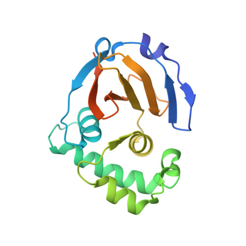
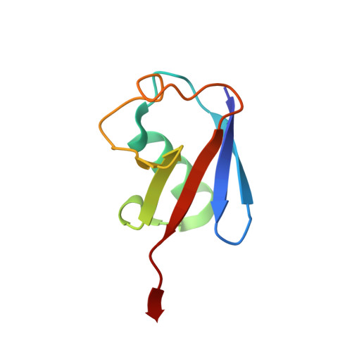
Structure of the Hazara virus OTU bound to ubiquitin


Experimental Data Snapshot

  • Method: X-RAY DIFFRACTION
  • Resolution: 2.78 Å
  • R-Value Free: 0.273 
  • R-Value Work: 0.235 
  • R-Value Observed: 0.237 

Starting Models: experimental
View more details

wwPDB Validation   3D Report Full Report


This is version 1.2 of the entry. See complete history


Literature

Flipping the substrate preference of Hazara virus ovarian tumour domain protease through structure-based mutagenesis.

Dzimianski, J.V.Mace, S.L.Williams, I.L.Freitas, B.T.Pegan, S.D.

(2020) Acta Crystallogr D Struct Biol 76: 1114-1123

  • DOI: https://doi.org/10.1107/S2059798320012875
  • Primary Citation of Related Structures:  
    7JMS

  • PubMed Abstract: 

    Nairoviruses are arthropod-borne viruses with a nearly global geographical distribution. Several are known causative agents of human disease, including Crimean-Congo hemorrhagic fever virus (CCHFV), which has a case fatality rate that can exceed 30%. Nairoviruses encode an ovarian tumour domain protease (OTU) that can suppress the innate immune response by reversing post-translational modifications by ubiquitin (Ub) and/or interferon-stimulated gene product 15 (ISG15). As a result, the OTU has been identified as a potential target for the development of CCHFV therapeutics. Despite sharing the same general fold, nairoviral OTUs show structural and enzymatic diversity. The CCHFV OTU, for example, possesses activity towards both Ub and ISG15, while the Hazara virus (HAZV) OTU interacts exclusively with Ub. Virology studies focused on the OTU have mostly been restricted to CCHFV, which requires BSL-4 containment facilities. Although HAZV has been proposed as a BSL-2 alternative, differences in the engagement of substrates by CCHFV and HAZV OTUs may present complicating factors when trying to model one using the other. To understand the molecular underpinnings of the differences in activity, a 2.78 Å resolution crystal structure of HAZV OTU bound to Ub was solved. Using structure-guided site-directed mutagenesis, HAZV OTUs were engineered with altered or eliminated deubiquitinase activity, including one with an exclusive activity for ISG15. Additionally, analysis of the structure yielded insights into the difference in inhibition observed between CCHFV and HAZV OTUs with a Ub-based inhibitor. These new insights present opportunities to utilize HAZV as a model system to better understand the role of the OTU in the context of infection.


  • Organizational Affiliation

    Pharmaceutical and Biomedical Sciences, University of Georgia, 240 West Green Street, Athens, GA 30602, USA.


Macromolecules
Find similar proteins by:  (by identity cutoff)  |  3D Structure
Entity ID: 1
MoleculeChains Sequence LengthOrganismDetailsImage
Replicase
A, C, E, G
178Orthonairovirus hazaraenseMutation(s): 0 
EC: 2.7.7.48 (PDB Primary Data), 3.4.19.12 (PDB Primary Data)
UniProt
Find proteins for A6XA53 (Hazara virus)
Explore A6XA53 
Go to UniProtKB:  A6XA53
Entity Groups  
Sequence Clusters30% Identity50% Identity70% Identity90% Identity95% Identity100% Identity
UniProt GroupA6XA53
Sequence Annotations
Expand
  • Reference Sequence
Find similar proteins by:  (by identity cutoff)  |  3D Structure
Entity ID: 2
MoleculeChains Sequence LengthOrganismDetailsImage
Polyubiquitin-B
B, D, F, H
75Homo sapiensMutation(s): 0 
Gene Names: UBB
UniProt & NIH Common Fund Data Resources
Find proteins for P0CG47 (Homo sapiens)
Explore P0CG47 
Go to UniProtKB:  P0CG47
PHAROS:  P0CG47
GTEx:  ENSG00000170315 
Entity Groups  
Sequence Clusters30% Identity50% Identity70% Identity90% Identity95% Identity100% Identity
UniProt GroupP0CG47
Sequence Annotations
Expand
  • Reference Sequence
Small Molecules
Ligands 3 Unique
IDChains Name / Formula / InChI Key2D Diagram3D Interactions
GOL
Query on GOL

Download Ideal Coordinates CCD File 
M [auth B],
P [auth C]
GLYCEROL
C3 H8 O3
PEDCQBHIVMGVHV-UHFFFAOYSA-N
AYE
Query on AYE

Download Ideal Coordinates CCD File 
L [auth B],
R [auth D],
U [auth F],
W [auth H]
prop-2-en-1-amine
C3 H7 N
VVJKKWFAADXIJK-UHFFFAOYSA-N
CA
Query on CA

Download Ideal Coordinates CCD File 
I [auth A]
J [auth A]
K [auth A]
N [auth C]
O [auth C]
I [auth A],
J [auth A],
K [auth A],
N [auth C],
O [auth C],
Q [auth D],
S [auth E],
T [auth F],
V [auth G]
CALCIUM ION
Ca
BHPQYMZQTOCNFJ-UHFFFAOYSA-N
Experimental Data & Validation

Experimental Data

  • Method: X-RAY DIFFRACTION
  • Resolution: 2.78 Å
  • R-Value Free: 0.273 
  • R-Value Work: 0.235 
  • R-Value Observed: 0.237 
  • Space Group: P 1 21 1
Unit Cell:
Length ( Å )Angle ( ˚ )
a = 83.85α = 90
b = 55.595β = 97.203
c = 97.434γ = 90
Software Package:
Software NamePurpose
PHENIXrefinement
HKL-2000data reduction
HKL-2000data scaling
PHENIXphasing

Structure Validation

View Full Validation Report


Entry History & Funding Information

Deposition Data


Funding OrganizationLocationGrant Number
National Institutes of Health/National Institute Of Allergy and Infectious Diseases (NIH/NIAID)United StatesR01AI109008

Revision History  (Full details and data files)

  • Version 1.0: 2020-10-21
    Type: Initial release
  • Version 1.1: 2020-11-18
    Changes: Database references
  • Version 1.2: 2023-10-18
    Changes: Data collection, Database references, Refinement description