Perturbative diffraction methods resolve a conformational switch that facilitates a two-step enzymatic mechanism.
Greisman, J.B., Dalton, K.M., Brookner, D.E., Klureza, M.A., Sheehan, C.J., Kim, I.S., Henning, R.W., Russi, S., Hekstra, D.R.(2024) Proc Natl Acad Sci U S A 121: e2313192121-e2313192121
- PubMed: 38386706 Search on PubMedSearch on PubMed Central
- DOI: https://doi.org/10.1073/pnas.2313192121
- Primary Citation Related Structures:
5SSS, 5SST, 5SSU, 5SSV, 5SSW, 7FPL, 7FPM, 7FPN, 7FPO, 7FPP, 7FPQ, 7FPR, 7FPS, 7FPT, 7FPU, 7FPV, 7FPW, 7FPX, 7FPY, 7FPZ, 7FQ0, 7FQ1, 7FQ2, 7FQ3, 7FQ4, 7FQ5, 7FQ6, 7FQ7, 7FQ8, 7FQ9, 7FQA, 7FQB, 7FQC, 7FQD, 7FQE, 7FQF, 7FQG, 8DAI, 8G4Z, 8G50 - PubMed Abstract:
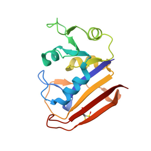
Enzymes catalyze biochemical reactions through precise positioning of substrates, cofactors, and amino acids to modulate the transition-state free energy. However, the role of conformational dynamics remains poorly understood due to poor experimental access. This shortcoming is evident with Escherichia coli dihydrofolate reductase (DHFR), a model system for the role of protein dynamics in catalysis, for which it is unknown how the enzyme regulates the different active site environments required to facilitate proton and hydride transfer. Here, we describe ligand-, temperature-, and electric-field-based perturbations during X-ray diffraction experiments to map the conformational dynamics of the Michaelis complex of DHFR. We resolve coupled global and local motions and find that these motions are engaged by the protonated substrate to promote efficient catalysis. This result suggests a fundamental design principle for multistep enzymes in which pre-existing dynamics enable intermediates to drive rapid electrostatic reorganization to facilitate subsequent chemical steps.
- Department of Molecular & Cellular Biology, Harvard University, Cambridge, MA 02138.
Organizational Affiliation: