Architecture of Dispatched, a Transmembrane Protein Responsible for Hedgehog Release.
Luo, Y., Wan, G., Zhou, X., Wang, Q., Zhang, Y., Bao, J., Cong, Y., Zhao, Y., Li, D.(2021) Front Mol Biosci 8: 701826-701826
- PubMed: 34557519 Search on PubMedSearch on PubMed Central
- DOI: https://doi.org/10.3389/fmolb.2021.701826
- Primary Citation Related Structures:
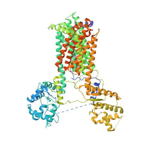
7FIF - PubMed Abstract:
The evolutionarily conserved Hedgehog (Hh) signaling pathway is crucial for programmed cell differentiation and proliferation. Dispatched (Disp) is a 12-transmembrane protein that plays a critical role in the Hedgehog (Hh) signaling pathway by releasing the dually lipidated ligand HhN from the membrane, a prerequisite step to the downstream signaling cascade. In this study, we focus on the Disp from water bear, a primitive animal known as the most indestructible on Earth. Using a zebrafish model, we show that the water bear homolog possesses the function of Disp. We have solved its structure to a 6.5-Å resolution using single-particle cryogenic electron microscopy. Consistent with the evolutional conservation of the pathway, the water bear Disp structure is overall similar to the previously reported structures of the fruit fly and human homologs. Although not revealing much detail at this resolution, the water bear Disp shows a different conformation compared to published structures, suggesting that they represent different functional snapshots.
- CAS Center for Excellence in Molecular Cell Science, Shanghai Institute of Biochemistry and Cell Biology, University of Chinese Academy of Sciences, Chinese Academy of Sciences, Shanghai, China.
Organizational Affiliation: