7FGX | pdb_00007fgx

Toxoplasma gondii dihydrofolate reductase thymidylate synthase (TgDHFR-TS) complexed with P39, NADPH and dUMP


Experimental Data Snapshot

  • Method: X-RAY DIFFRACTION
  • Resolution: 2.05 Å
  • R-Value Free: 
    0.214 (Depositor), 0.213 (DCC) 
  • R-Value Work: 
    0.180 (Depositor), 0.180 (DCC) 
  • R-Value Observed: 
    0.182 (Depositor) 

Starting Model: experimental
View more details

wwPDB Validation 3D Report Full Report

Validation slider image for 7FGX

Ligand Structure Quality Assessment 


This is version 1.3 of the entry. See complete history

Literature

Structural Insight into Effective Inhibitors' Binding to Toxoplasma gondii Dihydrofolate Reductase Thymidylate Synthase.

Vanichtanankul, J.Yoomuang, A.Taweechai, S.Saeyang, T.Pengon, J.Yuvaniyama, J.Tarnchompoo, B.Yuthavong, Y.Kamchonwongpaisan, S.

(2022) ACS Chem Biol 17: 1691-1702

  • DOI: https://doi.org/10.1021/acschembio.1c00627
  • Primary Citation Related Structures: 
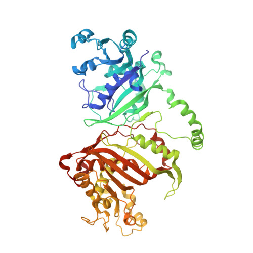
    7FGW, 7FGX, 7FGY, 7XI7

  • PubMed Abstract: 

    Pyrimethamine (Pyr), a known dihydrofolate reductase (DHFR) inhibitor, has long been used to treat toxoplasmosis caused by Toxoplasma gondii (Tg) infection. However, Pyr is effective only at high doses with associated toxicity to patients, calling for safer alternative treatments. In this study, we investigated a series of Pyr analogues, previously developed as DHFR inhibitors of Plasmodium falciparum bifunctional DHFR-thymidylate synthase (PfDHFR-TS), for their activity against T. gondii DHFR-TS (TgDHFR-TS). Of these, a set of compounds with a substitution at the C 6 position of the pyrimidine ring exhibited high binding affinities (in a low nanomolar range) against TgDHFR-TS and in vitro T. gondii inhibitory activity. Three-dimensional structures of TgDHFR-TS reported here include the ternary complexes with Pyr, P39, or P40. A comparison of these structures showed the minor steric strain between the p -chlorophenyl group of Pyr and Thr83 of TgDHFR-TS. Such a conflict was relieved in the complexes with the two analogues, P39 and P40, explaining their highest binding affinities described herein. Moreover, these structures suggested that the hydrophobic environment in the active-site pocket could be used for drug design to increase the potency and selectivity of antifolate inhibitors. These findings would accelerate the development of new antifolate drugs to treat toxoplasmosis.


  • Organizational Affiliation
    • National Center for Genetic Engineering and Biotechnology (BIOTEC), National Science and Technology Development Agency (NSTDA), 113 Thailand Science Park, Khlong Nueng, Khlong Luang, Pathum Thani 12120, Thailand.

Macromolecule Content 

  • Total Structure Weight: 280.67 kDa 
  • Atom Count: 19,328 
  • Modeled Residue Count: 2,241 
  • Deposited Residue Count: 2,440 
  • Unique protein chains: 1

Macromolecules

Find similar proteins by:|  3D Structure
Entity ID: 1
MoleculeChains  Sequence LengthOrganismDetailsImage
Bifunctional dihydrofolate reductase-thymidylate synthase
A, B, C, D
610Toxoplasma gondiiMutation(s): 0 
EC: 1.5.1.3 (PDB Primary Data), 2.1.1.45 (PDB Primary Data)
UniProt
Find proteins for Q07422 (Toxoplasma gondii)
Explore Q07422 
Go to UniProtKB:  Q07422
Entity Groups
Sequence Clusters30% Identity50% Identity70% Identity90% Identity95% Identity100% Identity
UniProt GroupQ07422
Sequence Annotations
Expand
Reference Sequence

Small Molecules

Ligands 3 Unique
IDChains Name / Formula / InChI Key2D Diagram3D Interactions
NDP

Query on NDP


Download:Ideal Coordinates CCD File
E [auth A],
H [auth B],
K [auth C],
N [auth D]
NADPH DIHYDRO-NICOTINAMIDE-ADENINE-DINUCLEOTIDE PHOSPHATE
C21 H30 N7 O17 P3
ACFIXJIJDZMPPO-NNYOXOHSSA-N
UMP

Query on UMP


Download:Ideal Coordinates CCD File
G [auth A],
J [auth B],
M [auth C],
P [auth D]
2'-DEOXYURIDINE 5'-MONOPHOSPHATE
C9 H13 N2 O8 P
JSRLJPSBLDHEIO-SHYZEUOFSA-N
4RI
(Subject of Investigation/LOI)

Query on 4RI


Download:Ideal Coordinates CCD File
F [auth A],
I [auth B],
L [auth C],
O [auth D]
6-hexyl-5-phenyl-pyrimidine-2,4-diamine
C16 H22 N4
DBCDYKFZHWOABJ-UHFFFAOYSA-N

Experimental Data & Validation

Experimental Data

  • Method: X-RAY DIFFRACTION
  • Resolution: 2.05 Å
  • R-Value Free:  0.214 (Depositor), 0.213 (DCC) 
  • R-Value Work:  0.180 (Depositor), 0.180 (DCC) 
  • R-Value Observed: 0.182 (Depositor) 
Space Group: P 1
Unit Cell:
Length ( Å )Angle ( ˚ )
a = 68.404α = 88.95
b = 102.536β = 87.74
c = 106.198γ = 84.69
Software Package:
Software NamePurpose
PHENIXrefinement
PHENIXrefinement
PHASERphasing
PHASERphasing
HKL-2000data reduction
HKL-2000data scaling
PHASERphasing

Structure Validation

View Full Validation Report


Ligand Structure Quality Assessment 


Entry History 

& Funding Information

Deposition Data


Funding OrganizationLocationGrant Number
The Thailand Research Fund (TRF)ThailandTRG5780138

Revision History  (Full details and data files)

  • Version 1.0: 2022-06-29
    Type: Initial release
  • Version 1.1: 2022-07-06
    Changes: Derived calculations
  • Version 1.2: 2022-07-27
    Changes: Database references
  • Version 1.3: 2023-11-29
    Changes: Data collection, Refinement description