7FFP | pdb_00007ffp

Crystal structure of di-peptidase-E from Xenopus laevis


Experimental Data Snapshot

  • Method: X-RAY DIFFRACTION
  • Resolution: 1.80 Å
  • R-Value Free: 
    0.223 (Depositor), 0.221 (DCC) 
  • R-Value Work: 
    0.187 (Depositor), 0.186 (DCC) 
  • R-Value Observed: 
    0.188 (Depositor) 

Starting Model: experimental
View more details

wwPDB Validation 3D Report Full Report

Validation slider image for 7FFP

Ligand Structure Quality Assessment 


This is version 1.2 of the entry. See complete history

Literature

Crystal structure of aspartyl dipeptidase from Xenopus laevis revealed ligand binding induced loop ordering and catalytic triad assembly.

Kumar, A.Singh, R.Ghosh, B.Makde, R.D.

(2022) Proteins 90: 299-308

  • DOI: https://doi.org/10.1002/prot.26220
  • Primary Citation Related Structures: 
    7C9B, 7FFP

  • PubMed Abstract: 

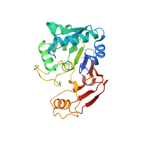
    Gene encoding aspartyl dipeptidase from Xenopus levies (PepExl) is upregulated by thyroid hormone and is proposed to play a significant role in resorption of tadpole tail during metamorphosis. However, the importance of peptidase activity for the resorption of the tail remain elusive. Here we report the crystal structures of first eukaryotic S51 peptidase, PepExl, in its ligand-free and Asp-bound states at 1.4 and 1.8 Å resolutions, respectively. The active site is located at dimeric interface and the catalytic triad is found to be dissembled in ligand-free and assembled in Asp-bound state. Structural comparison and molecular dynamic simulations of ligand-free and Asp-bound states shows that distinct loop (loop-A) plays an important role in active site shielding, substrate binding and enzyme activation. This study illuminates the Asp-X dipeptide binding in PepExl is associated with ordering of the loop-A and assembly of residues of catalytic triad in active conformation for enzymatic activity.


  • Organizational Affiliation
    • Beamline Development and Application Section, Bhabha Atomic Research Centre, Mumbai, India.

Macromolecule Content 

  • Total Structure Weight: 27.16 kDa 
  • Atom Count: 2,126 
  • Modeled Residue Count: 240 
  • Deposited Residue Count: 242 
  • Unique protein chains: 1

Macromolecules

Find similar proteins by:|  3D Structure
Entity ID: 1
MoleculeChains  Sequence LengthOrganismDetailsImage
Alpha-aspartyl dipeptidase242Xenopus laevisMutation(s): 0 
Gene Names: aad-a
EC: 3.4.13.21
UniProt
Find proteins for Q91642 (Xenopus laevis)
Explore Q91642 
Go to UniProtKB:  Q91642
Entity Groups
Sequence Clusters30% Identity50% Identity70% Identity90% Identity95% Identity100% Identity
UniProt GroupQ91642
Sequence Annotations
Expand
Reference Sequence

Experimental Data & Validation

Experimental Data

  • Method: X-RAY DIFFRACTION
  • Resolution: 1.80 Å
  • R-Value Free:  0.223 (Depositor), 0.221 (DCC) 
  • R-Value Work:  0.187 (Depositor), 0.186 (DCC) 
  • R-Value Observed: 0.188 (Depositor) 
Space Group: C 1 2 1
Unit Cell:
Length ( Å )Angle ( ˚ )
a = 82.666α = 90
b = 42.15β = 118.24
c = 75.68γ = 90
Software Package:
Software NamePurpose
XDSdata reduction
Aimlessdata scaling
PHASERphasing
PHENIXrefinement
PDB_EXTRACTdata extraction

Structure Validation

View Full Validation Report


Ligand Structure Quality Assessment 


Entry History 

Deposition Data

Revision History  (Full details and data files)

  • Version 1.0: 2021-09-08
    Type: Initial release
  • Version 1.1: 2022-02-16
    Changes: Database references
  • Version 1.2: 2023-11-29
    Changes: Data collection, Refinement description