7FFM | pdb_00007ffm

Human serum transferrin with five osmium binding sites

  • Classification: METAL TRANSPORT
  • Organism(s): Homo sapiens
  • Mutation(s): Yes 

  • Deposited: 2021-07-23 Released: 2022-06-22 
  • Deposition Author(s): Wang, M., Sun, H.
  • Funding Organization(s): The University Grants Committee, Research Grants Council (RGC), National Science Foundation (NSF, China)

Experimental Data Snapshot

  • Method: X-RAY DIFFRACTION
  • Resolution: 3.06 Å
  • R-Value Free: 
    0.212 (Depositor), 0.212 (DCC) 
  • R-Value Work: 
    0.179 (Depositor), 0.181 (DCC) 
  • R-Value Observed: 
    0.180 (Depositor) 

Starting Model: experimental
View more details

wwPDB Validation 3D Report Full Report

Validation slider image for 7FFM

This is version 2.2 of the entry. See complete history

Literature

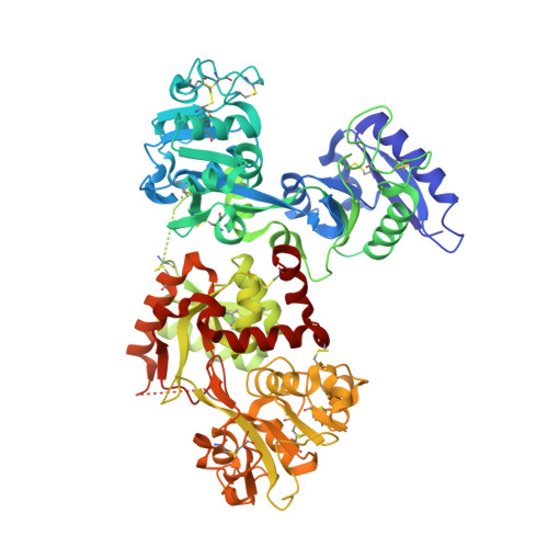
Binding of ruthenium and osmium at non‐iron sites of transferrin accounts for their iron-independent cellular uptake.

Wang, M.Wang, H.Xu, X.Lai, T.P.Zhou, Y.Hao, Q.Li, H.Sun, H.

(2022) J Inorg Biochem 234: 111885-111885

  • DOI: https://doi.org/10.1016/j.jinorgbio.2022.111885
  • Primary Citation Related Structures: 
    5WTD, 5X5P, 7FFM, 7FFU

  • PubMed Abstract: 

    Being identified with less toxic and generally showing selective effects for solid tumor metastases, ruthenium and osmium compounds are promising drug candidates for clinical uses. Human serum proteins, such as albumin and transferrin, play vital roles in the transportation and accumulation of ruthenium and osmium agents into target tissues. However, the molecular mechanism of how transferrin transport ruthenium and their osmium analogues at atomic level remains obscure. In this study, we uncovered that the cellular uptake of Os 3+ or Ru 3+ are not competed by Fe 3+ . To unveil the molecular mechanism behind the phenomena, we report the first crystal structures of human serum transferrin (hTF) in complex with ruthenium and osmium compounds bound to the non-conserved residues on the surface of hTF without altering its overall conformation. As for Ru 3+ and Os 3+ , these binding sites by descending affinity are: His14/His289, His349-350 ~ His578/Arg581. Ruthenium drugs and their osmium analogues preferentially bind to His14/His289 with bipyridine or imidazole ligands leaving. These binding sites on hTF surface are also available in human lactoferrin and some transferrin family member of other species. The presence of these binding sites makes the cellular uptake of Ru 3+ and Os 3+ less affected by Fe 3+ , compare to Zr 4+ or Hf 4+ . Collectively, these findings are critical for our understanding of the role of serum transferrin in cellular delivery of ruthenium and osmium anticancer agents.


  • Organizational Affiliation
    • Department of Chemistry and CAS-HKU Joint Laboratory of Metallomics for Health and Environment, the University of Hong Kong, Pokfulam Road, Hong Kong SAR, China; School of Chemistry and Molecular Engineering, East China Normal University, No. 3663 Zhong Shan Road North, Shanghai 200062, China.

Macromolecule Content 

  • Total Structure Weight: 76.58 kDa 
  • Atom Count: 5,044 
  • Modeled Residue Count: 649 
  • Deposited Residue Count: 679 
  • Unique protein chains: 1

Macromolecules

Find similar proteins by:|  3D Structure
Entity ID: 1
MoleculeChains  Sequence LengthOrganismDetailsImage
Serotransferrin679Homo sapiensMutation(s): 1 
UniProt & NIH Common Fund Data Resources
Find proteins for P02787 (Homo sapiens)
Explore P02787 
Go to UniProtKB:  P02787
PHAROS:  P02787
GTEx:  ENSG00000091513 
Entity Groups
Sequence Clusters30% Identity50% Identity70% Identity90% Identity95% Identity100% Identity
UniProt GroupP02787
Sequence Annotations
Expand
Reference Sequence

Small Molecules

Ligands 4 Unique
IDChains Name / Formula / InChI Key2D Diagram3D Interactions
NTA

Query on NTA


Download:Ideal Coordinates CCD File
D [auth A]NITRILOTRIACETIC ACID
C6 H9 N O6
MGFYIUFZLHCRTH-UHFFFAOYSA-N
OS
(Subject of Investigation/LOI)

Query on OS


Download:Ideal Coordinates CCD File
E [auth A],
F [auth A],
G [auth A],
H [auth A],
I [auth A]
OSMIUM ION
Os
XQBKHDFIPARBOX-UHFFFAOYSA-N
MLI

Query on MLI


Download:Ideal Coordinates CCD File
C [auth A]MALONATE ION
C3 H2 O4
OFOBLEOULBTSOW-UHFFFAOYSA-L
4TI

Query on 4TI


Download:Ideal Coordinates CCD File
B [auth A]TITANIUM ION
Ti
VEOOXLFYKHRLHJ-UHFFFAOYSA-N

Experimental Data & Validation

Experimental Data

  • Method: X-RAY DIFFRACTION
  • Resolution: 3.06 Å
  • R-Value Free:  0.212 (Depositor), 0.212 (DCC) 
  • R-Value Work:  0.179 (Depositor), 0.181 (DCC) 
  • R-Value Observed: 0.180 (Depositor) 
Space Group: C 2 2 21
Unit Cell:
Length ( Å )Angle ( ˚ )
a = 137.954α = 90
b = 158.211β = 90
c = 107.102γ = 90
Software Package:
Software NamePurpose
SCALEPACKdata scaling
PHENIXrefinement
PDB_EXTRACTdata extraction
HKL-2000data reduction
PHASERphasing

Structure Validation

View Full Validation Report


Entry History 

& Funding Information

Deposition Data


Funding OrganizationLocationGrant Number
The University Grants Committee, Research Grants Council (RGC)Hong Kong1733616P
National Science Foundation (NSF, China)China21671203

Revision History  (Full details and data files)

  • Version 1.0: 2022-06-22
    Type: Initial release
  • Version 2.0: 2023-11-15
    Changes: Atomic model, Data collection, Derived calculations
  • Version 2.1: 2023-11-29
    Changes: Refinement description
  • Version 2.2: 2024-11-13
    Changes: Structure summary