


Non-Specific Class-c acidphosphatase from Sphingobium sp. RSMS
Gaur, N.K., Kumar, A., Sunder, S., Mukhopadhyaya, R., Makde, R.D.To be published.
Experimental Data Snapshot
Entity ID: 1 | |||||
|---|---|---|---|---|---|
| Molecule | Chains | Sequence Length | Organism | Details | Image |
| Acid phosphatase | 296 | Sphingobium sp. 20006FA | Mutation(s): 0 Gene Names: A8O16_10785 | 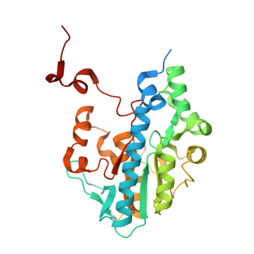 | |
| Ligands 4 Unique | |||||
|---|---|---|---|---|---|
| ID | Chains | Name / Formula / InChI Key | 2D Diagram | 3D Interactions | |
| ADN (Subject of Investigation/LOI) Download:Ideal Coordinates CCD File | B [auth A] | ADENOSINE C10 H13 N5 O4 OIRDTQYFTABQOQ-KQYNXXCUSA-N |  | ||
| PEG Download:Ideal Coordinates CCD File | C [auth A] | DI(HYDROXYETHYL)ETHER C4 H10 O3 MTHSVFCYNBDYFN-UHFFFAOYSA-N |  | ||
| PO4 (Subject of Investigation/LOI) Download:Ideal Coordinates CCD File | E [auth A] | PHOSPHATE ION O4 P NBIIXXVUZAFLBC-UHFFFAOYSA-K |  | ||
| MG (Subject of Investigation/LOI) Download:Ideal Coordinates CCD File | D [auth A] | MAGNESIUM ION Mg JLVVSXFLKOJNIY-UHFFFAOYSA-N |  | ||
| Length ( Å ) | Angle ( ˚ ) |
|---|---|
| a = 167.937 | α = 90 |
| b = 167.937 | β = 90 |
| c = 167.937 | γ = 90 |
| Software Name | Purpose |
|---|---|
| PHENIX | refinement |
| PDB_EXTRACT | data extraction |
| XDS | data reduction |
| Aimless | data scaling |
| MR-Rosetta | phasing |


