7F1M

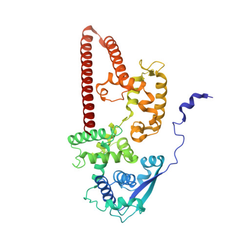
Marburg virus nucleoprotein-RNA complex


Experimental Data Snapshot

  • Method: ELECTRON MICROSCOPY
  • Resolution: 3.10 Å
  • Aggregation State: HELICAL ARRAY 
  • Reconstruction Method: SINGLE PARTICLE 

Starting Model: experimental
View more details

wwPDB Validation   3D Report Full Report


This is version 1.2 of the entry. See complete history


Literature

Structural insight into Marburg virus nucleoprotein-RNA complex formation.

Fujita-Fujiharu, Y.Sugita, Y.Takamatsu, Y.Houri, K.Igarashi, M.Muramoto, Y.Nakano, M.Tsunoda, Y.Taniguchi, I.Becker, S.Noda, T.

(2022) Nat Commun 13: 1191-1191

  • DOI: https://doi.org/10.1038/s41467-022-28802-x
  • Primary Citation of Related Structures:  
    7F1M

  • PubMed Abstract: 

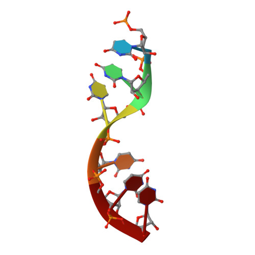
    The nucleoprotein (NP) of Marburg virus (MARV), a close relative of Ebola virus (EBOV), encapsidates the single-stranded, negative-sense viral genomic RNA (vRNA) to form the helical NP-RNA complex. The NP-RNA complex constitutes the core structure for the assembly of the nucleocapsid that is responsible for viral RNA synthesis. Although appropriate interactions among NPs and RNA are required for the formation of nucleocapsid, the structural basis of the helical assembly remains largely elusive. Here, we show the structure of the MARV NP-RNA complex determined using cryo-electron microscopy at a resolution of 3.1 Å. The structures of the asymmetric unit, a complex of an NP and six RNA nucleotides, was very similar to that of EBOV, suggesting that both viruses share common mechanisms for the nucleocapsid formation. Structure-based mutational analysis of both MARV and EBOV NPs identified key residues for helical assembly and subsequent viral RNA synthesis. Importantly, most of the residues identified were conserved in both viruses. These findings provide a structural basis for understanding the nucleocapsid formation and contribute to the development of novel antivirals against MARV and EBOV.


  • Organizational Affiliation

    Laboratory of Ultrastructural Virology, Institute for Frontier Life and Medical Sciences, Kyoto University, 53 Shogoin Kawahara-cho, Sakyo-ku, Kyoto, 606-8507, Japan.


Macromolecules

Find similar proteins by:  (by identity cutoff)  |  3D Structure
Entity ID: 1
MoleculeChains Sequence LengthOrganismDetailsImage
NucleoproteinA,
C [auth B]
395Lake Victoria marburgvirus - Angola2005Mutation(s): 2 
Gene Names: NP
UniProt
Find proteins for Q1PD53 (Lake Victoria marburgvirus (strain Angola/2005))
Explore Q1PD53 
Go to UniProtKB:  Q1PD53
Entity Groups  
Sequence Clusters30% Identity50% Identity70% Identity90% Identity95% Identity100% Identity
UniProt GroupQ1PD53
Sequence Annotations
Expand
  • Reference Sequence

Find similar nucleic acids by:  Sequence   |   3D Structure  

Entity ID: 2
MoleculeChains LengthOrganismImage
RNA (5'-R(P*UP*UP*UP*UP*UP*U)-3')B [auth R],
D [auth S]
6synthetic construct
Sequence Annotations
Expand
  • Reference Sequence
Experimental Data & Validation

Experimental Data

  • Method: ELECTRON MICROSCOPY
  • Resolution: 3.10 Å
  • Aggregation State: HELICAL ARRAY 
  • Reconstruction Method: SINGLE PARTICLE 
EM Software:
TaskSoftware PackageVersion
RECONSTRUCTIONRELION3.1.0
MODEL REFINEMENTPHENIX1.18

Structure Validation

View Full Validation Report


Entry History & Funding Information

Deposition Data


Funding OrganizationLocationGrant Number
Japan Society for the Promotion of Science (JSPS)Japan20H03494
Japan Society for the Promotion of Science (JSPS)Japan19K22529
Japan Society for the Promotion of Science (JSPS)Japan19H04831
Japan Agency for Medical Research and Development (AMED)Japan19fk0108113
Japan Agency for Medical Research and Development (AMED)Japan20fk0108270h0001
Japan Science and TechnologyJapanJPMJCR20HA

Revision History  (Full details and data files)

  • Version 1.0: 2022-03-09
    Type: Initial release
  • Version 1.1: 2022-04-06
    Changes: Database references
  • Version 1.2: 2024-06-12
    Changes: Data collection, Refinement description