7EZY | pdb_00007ezy

anti-CRISPR-associated Aca2


Experimental Data Snapshot

  • Method: X-RAY DIFFRACTION
  • Resolution: 1.92 Å
  • R-Value Free: 
    0.232 (Depositor), 0.214 (DCC) 
  • R-Value Work: 
    0.198 (Depositor) 
  • R-Value Observed: 
    0.201 (Depositor) 

Starting Model: experimental
View more details

wwPDB Validation 3D Report Full Report

Validation slider image for 7EZY

This is version 1.2 of the entry. See complete history

Literature

Molecular basis of transcriptional repression of anti-CRISPR by anti-CRISPR-associated 2.

Lee, S.Y.Kim, G.E.Park, H.H.

(2022) Acta Crystallogr D Struct Biol 78: 59-68

  • DOI: https://doi.org/10.1107/S2059798321011670
  • Primary Citation Related Structures: 
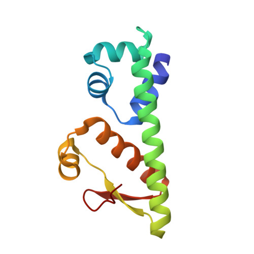
    7EZY

  • PubMed Abstract: 

    CRISPR-Cas systems are well known host defense mechanisms that are conserved in bacteria and archaea. To counteract CRISPR-Cas systems, phages and viruses have evolved to possess multiple anti-CRISPR (Acr) proteins that can inhibit the host CRISPR-Cas system via different strategies. The expression of acr genes is controlled by anti-CRISPR-associated (Aca) proteins that bind to an upstream promoter and regulate the expression of acr genes during transcription. Although the role of Aca as a transcriptional repressor has been demonstrated, the mechanism of action of Aca has not been determined. Here, the molecular mechanism underlying the Aca2-mediated transcriptional control of acr genes was elucidated by determining the crystal structure of Aca2 from Oceanimonas smirnovii at a high resolution of 1.92 Å. Aca2 forms a dimer in solution, and dimerization of Aca2 is critical for specific promoter binding. The promoter-binding strategy of dimeric Aca2 was also revealed by performing mutagenesis studies. The atomic structure of the Aca family shown in this study provides insights into the fine regulation of host defense and immune-escape mechanisms and also demonstrates the conserved working mechanism of the Aca family.


  • Organizational Affiliation
    • College of Pharmacy, Chung-Ang University, Seoul 06974, Republic of Korea.

Macromolecule Content 

  • Total Structure Weight: 28.66 kDa 
  • Atom Count: 2,184 
  • Modeled Residue Count: 248 
  • Deposited Residue Count: 250 
  • Unique protein chains: 1

Macromolecules

Find similar proteins by:|  3D Structure
Entity ID: 1
MoleculeChains  Sequence LengthOrganismDetailsImage
anti-CRISPR-associated Aca2
A, B
125Oceanimonas smirnoviiMutation(s): 0 
UniProt
Find proteins for A0A8M0FGS8 (Oceanimonas smirnovii)
Explore A0A8M0FGS8 
Go to UniProtKB:  A0A8M0FGS8
Entity Groups
Sequence Clusters30% Identity50% Identity70% Identity90% Identity95% Identity100% Identity
UniProt GroupA0A8M0FGS8
Sequence Annotations
Expand
Reference Sequence

Experimental Data & Validation

Experimental Data

  • Method: X-RAY DIFFRACTION
  • Resolution: 1.92 Å
  • R-Value Free:  0.232 (Depositor), 0.214 (DCC) 
  • R-Value Work:  0.198 (Depositor) 
  • R-Value Observed: 0.201 (Depositor) 
Space Group: P 41 21 2
Unit Cell:
Length ( Å )Angle ( ˚ )
a = 64.13α = 90
b = 64.13β = 90
c = 135.52γ = 90
Software Package:
Software NamePurpose
PHENIXrefinement
PHENIXrefinement
XDSdata reduction
XDSdata scaling
PHENIXphasing

Structure Validation

View Full Validation Report


Entry History 

& Funding Information

Deposition Data


Funding OrganizationLocationGrant Number
National Research Foundation (NRF, Korea)Korea, Republic Of--

Revision History  (Full details and data files)

  • Version 1.0: 2022-01-12
    Type: Initial release
  • Version 1.1: 2023-11-29
    Changes: Data collection, Refinement description
  • Version 1.2: 2025-08-06
    Changes: Database references, Structure summary