7EUO | pdb_00007euo

The structure of formyl peptide receptor 1 in complex with Gi and peptide agonist fMLF


Experimental Data Snapshot

  • Method: ELECTRON MICROSCOPY
  • Resolution: 2.90 Å
  • Aggregation State: PARTICLE 
  • Reconstruction Method: SINGLE PARTICLE 

Starting Model: experimental
View more details

wwPDB Validation 3D Report Full Report

Validation slider image for 7EUO

This is version 1.2 of the entry. See complete history

Literature

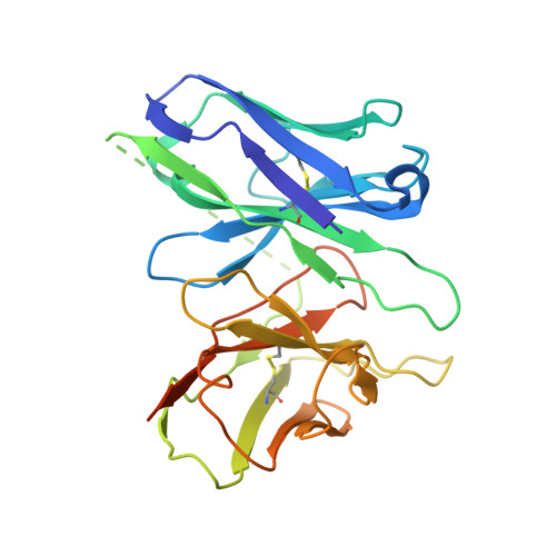
Structural basis for recognition of N-formyl peptides as pathogen-associated molecular patterns.

Chen, G.Wang, X.Liao, Q.Ge, Y.Jiao, H.Chen, Q.Liu, Y.Lyu, W.Zhu, L.van Zundert, G.C.P.Robertson, M.J.Skiniotis, G.Du, Y.Hu, H.Ye, R.D.

(2022) Nat Commun 13: 5232-5232

  • DOI: https://doi.org/10.1038/s41467-022-32822-y
  • Primary Citation Related Structures: 
    7EUO, 7VFX

  • PubMed Abstract: 

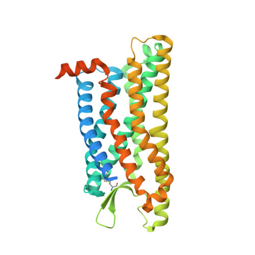
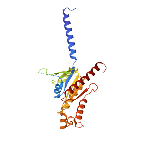
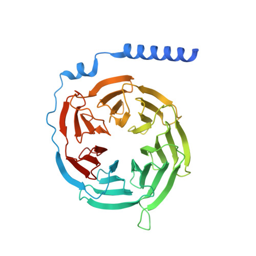
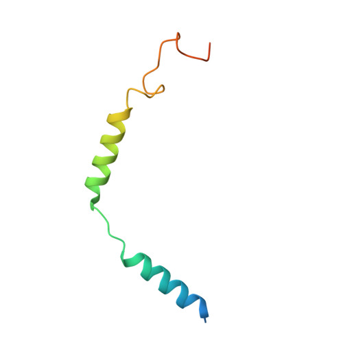
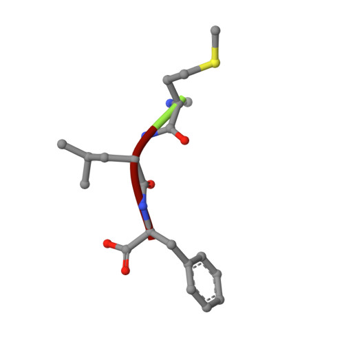
    The formyl peptide receptor 1 (FPR1) is primarily responsible for detection of short peptides bearing N-formylated methionine (fMet) that are characteristic of protein synthesis in bacteria and mitochondria. As a result, FPR1 is critical to phagocyte migration and activation in bacterial infection, tissue injury and inflammation. How FPR1 distinguishes between formyl peptides and non-formyl peptides remains elusive. Here we report cryo-EM structures of human FPR1-Gi protein complex bound to S. aureus-derived peptide fMet-Ile-Phe-Leu (fMIFL) and E. coli-derived peptide fMet-Leu-Phe (fMLF). Both structures of FPR1 adopt an active conformation and exhibit a binding pocket containing the R201 5.38 XXXR205 5.42 (RGIIR) motif for formyl group interaction and receptor activation. This motif works together with D106 3.33 for hydrogen bond formation with the N-formyl group and with fMet, a model supported by MD simulation and functional assays of mutant receptors with key residues for recognition substituted by alanine. The cryo-EM model of agonist-bound FPR1 provides a structural basis for recognition of bacteria-derived chemotactic peptides with potential applications in developing FPR1-targeting agents.


  • Organizational Affiliation
    • Kobilka Institute of Innovative Drug Discovery, School of Medicine, The Chinese University of Hong Kong, Shenzhen, Guangdong, 518172, China.

Macromolecule Content 

  • Total Structure Weight: 159.47 kDa 
  • Atom Count: 8,950 
  • Modeled Residue Count: 1,138 
  • Deposited Residue Count: 1,432 
  • Unique protein chains: 6

Macromolecules

Find similar proteins by:|  3D Structure
Entity ID: 1
MoleculeChains  Sequence LengthOrganismDetailsImage
fMet-Leu-Phe receptorA [auth R]378Homo sapiensMutation(s): 0 
Gene Names: FPR1
Membrane Entity: Yes 
UniProt & NIH Common Fund Data Resources
Find proteins for P21462 (Homo sapiens)
Explore P21462 
Go to UniProtKB:  P21462
PHAROS:  P21462
GTEx:  ENSG00000171051 
Entity Groups
Sequence Clusters30% Identity50% Identity70% Identity90% Identity95% Identity100% Identity
UniProt GroupP21462
Sequence Annotations
Expand
Reference Sequence
Find similar proteins by:|  3D Structure
Entity ID: 2
MoleculeChains  Sequence LengthOrganismDetailsImage
Guanine nucleotide-binding protein G(i) subunit alpha-1B [auth A]354Homo sapiensMutation(s): 0 
Gene Names: GNAI1
EC: 3.6.5
Membrane Entity: Yes 
UniProt & NIH Common Fund Data Resources
Find proteins for P63096 (Homo sapiens)
Explore P63096 
Go to UniProtKB:  P63096
PHAROS:  P63096
GTEx:  ENSG00000127955 
Entity Groups
Sequence Clusters30% Identity50% Identity70% Identity90% Identity95% Identity100% Identity
UniProt GroupP63096
Sequence Annotations
Expand
Reference Sequence
Find similar proteins by:|  3D Structure
Entity ID: 3
MoleculeChains  Sequence LengthOrganismDetailsImage
Guanine nucleotide-binding protein G(I)/G(S)/G(T) subunit beta-1C [auth B]357Homo sapiensMutation(s): 0 
Gene Names: GNB1
UniProt & NIH Common Fund Data Resources
Find proteins for P62873 (Homo sapiens)
Explore P62873 
Go to UniProtKB:  P62873
PHAROS:  P62873
GTEx:  ENSG00000078369 
Entity Groups
Sequence Clusters30% Identity50% Identity70% Identity90% Identity95% Identity100% Identity
UniProt GroupP62873
Sequence Annotations
Expand
Reference Sequence
Find similar proteins by:|  3D Structure
Entity ID: 4
MoleculeChains  Sequence LengthOrganismDetailsImage
Guanine nucleotide-binding protein G(I)/G(S)/G(O) subunit gamma-2D [auth G]71Homo sapiensMutation(s): 0 
Gene Names: GNG2
Membrane Entity: Yes 
UniProt & NIH Common Fund Data Resources
Find proteins for P59768 (Homo sapiens)
Explore P59768 
Go to UniProtKB:  P59768
PHAROS:  P59768
GTEx:  ENSG00000186469 
Entity Groups
Sequence Clusters30% Identity50% Identity70% Identity90% Identity95% Identity100% Identity
UniProt GroupP59768
Sequence Annotations
Expand
Reference Sequence
Find similar proteins by:|  3D Structure
Entity ID: 5
MoleculeChains  Sequence LengthOrganismDetailsImage
scFv16E [auth S]269Mus musculusMutation(s): 0 
Find similar proteins by:  Sequence   |   3D Structure  
Entity ID: 6
MoleculeChains  Sequence LengthOrganismDetailsImage
Peptide Agonist fMLFF [auth C]3Escherichia coliMutation(s): 0 

Small Molecules

Modified Residues  1 Unique
IDChains TypeFormula2D DiagramParent
FME
Query on FME
F [auth C]L-PEPTIDE LINKINGC6 H11 N O3 SMET

Experimental Data & Validation

Experimental Data

  • Method: ELECTRON MICROSCOPY
  • Resolution: 2.90 Å
  • Aggregation State: PARTICLE 
  • Reconstruction Method: SINGLE PARTICLE 
EM Software:
TaskSoftware PackageVersion
RECONSTRUCTIONcryoSPARC
MODEL REFINEMENTPHENIX

Structure Validation

View Full Validation Report


Entry History 

& Funding Information

Deposition Data


Funding OrganizationLocationGrant Number
Other government--

Revision History  (Full details and data files)

  • Version 1.0: 2022-05-25
    Type: Initial release
  • Version 1.1: 2022-12-07
    Changes: Database references, Refinement description
  • Version 1.2: 2024-11-06
    Changes: Data collection, Refinement description, Structure summary