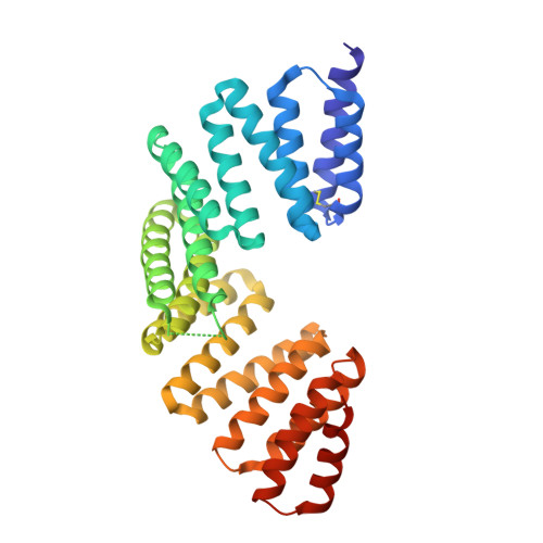
7EP7

The complex structure of Gpsm2 and Whirlin


Experimental Data Snapshot

  • Method: X-RAY DIFFRACTION
  • Resolution: 2.60 Å
  • R-Value Free: 0.283 
  • R-Value Work: 0.231 
  • R-Value Observed: 0.236 

Starting Model: experimental
View more details

wwPDB Validation   3D Report Full Report


This is version 1.3 of the entry. See complete history


Literature

Promotion of row 1-specific tip complex condensates by Gpsm2-G alpha i provides insights into row identity of the tallest stereocilia.

Shi, Y.Lin, L.Wang, C.Zhu, J.

(2022) Sci Adv 8: eabn4556-eabn4556

  • DOI: https://doi.org/10.1126/sciadv.abn4556
  • Primary Citation of Related Structures:  
    7EP7

  • PubMed Abstract: 

    The mechanosensory stereocilia in hair cells are organized into rows of graded height, a property crucial for auditory perception. Gpsm2-Gαi-Whirlin-Myo15-Eps8 complex at tips of the tallest stereocilia is proposed to define hair bundle row identity, although the underlying mechanism remains elusive. Here, we find that Gpsm2 could undergo phase separation. Moreover, row 1-specific Gpsm2-Gαi complex significantly promotes the formation of the five-component tip complex density (5xTCD) condensates. The 5xTCD condensates display much stronger actin-bundling ability than those without Gpsm2-Gαi, which may provide critical insights into how Gpsm2-Gαi specifies the tallest stereocilia. A deafness-associated mutation of Gpsm2 leads to impaired formation of the 5xTCD condensates and consequently reduced actin bundling, providing possible clues for etiology of hearing loss in patients with Chudley-McCullough syndrome.


  • Organizational Affiliation

    Department of Neurology, the First Affiliated Hospital of USTC, Ministry of Education Key Laboratory for Cellular Dynamics, Biomedical Sciences and Health Laboratory of Anhui Province, School of Life Sciences, Division of Life Sciences and Medicine, University of Science and Technology of China, Hefei 230027, China.


Macromolecules
Find similar proteins by:  (by identity cutoff)  |  3D Structure
Entity ID: 1
MoleculeChains Sequence LengthOrganismDetailsImage
G-protein-signaling modulator 2340Mus musculusMutation(s): 0 
Gene Names: Gpsm2LgnPins
UniProt & NIH Common Fund Data Resources
Find proteins for Q8VDU0 (Mus musculus)
Explore Q8VDU0 
Go to UniProtKB:  Q8VDU0
IMPC:  MGI:1923373
Entity Groups  
Sequence Clusters30% Identity50% Identity70% Identity90% Identity95% Identity100% Identity
UniProt GroupQ8VDU0
Sequence Annotations
Expand
  • Reference Sequence
Find similar proteins by:  (by identity cutoff)  |  3D Structure
Entity ID: 2
MoleculeChains Sequence LengthOrganismDetailsImage
Whirlin30Homo sapiensMutation(s): 0 
Gene Names: WHRNDFNB31KIAA1526
UniProt & NIH Common Fund Data Resources
Find proteins for Q9P202 (Homo sapiens)
Explore Q9P202 
Go to UniProtKB:  Q9P202
PHAROS:  Q9P202
GTEx:  ENSG00000095397 
Entity Groups  
Sequence Clusters30% Identity50% Identity70% Identity90% Identity95% Identity100% Identity
UniProt GroupQ9P202
Sequence Annotations
Expand
  • Reference Sequence
Experimental Data & Validation

Experimental Data

  • Method: X-RAY DIFFRACTION
  • Resolution: 2.60 Å
  • R-Value Free: 0.283 
  • R-Value Work: 0.231 
  • R-Value Observed: 0.236 
  • Space Group: P 61 2 2
Unit Cell:
Length ( Å )Angle ( ˚ )
a = 91.885α = 90
b = 91.885β = 90
c = 177.202γ = 120
Software Package:
Software NamePurpose
PHENIXrefinement
XSCALEdata scaling
PHASERphasing
PDB_EXTRACTdata extraction
XDSdata reduction

Structure Validation

View Full Validation Report


Entry History 

Deposition Data

Revision History  (Full details and data files)

  • Version 1.0: 2022-05-04
    Type: Initial release
  • Version 1.1: 2022-11-16
    Changes: Database references
  • Version 1.2: 2023-11-29
    Changes: Data collection, Refinement description
  • Version 1.3: 2024-11-06
    Changes: Structure summary