7EOP | pdb_00007eop

Crystal structure of the Pepper aptamer in complex with HBC620


Experimental Data Snapshot

  • Method: X-RAY DIFFRACTION
  • Resolution: 1.80 Å
  • R-Value Free: 
    0.211 (Depositor), 0.212 (DCC) 
  • R-Value Work: 
    0.190 (Depositor), 0.191 (DCC) 
  • R-Value Observed: 
    0.191 (Depositor) 

Starting Model: experimental
View more details

wwPDB Validation 3D Report Full Report

Validation slider image for 7EOP

Ligand Structure Quality Assessment 


This is version 1.2 of the entry. See complete history

Literature

Structure-based investigation of fluorogenic Pepper aptamer.

Huang, K.Chen, X.Li, C.Song, Q.Li, H.Zhu, L.Yang, Y.Ren, A.

(2021) Nat Chem Biol 17: 1289-1295

  • DOI: https://doi.org/10.1038/s41589-021-00884-6
  • Primary Citation Related Structures: 
    7EOG, 7EOH, 7EOI, 7EOJ, 7EOK, 7EOL, 7EOM, 7EON, 7EOO, 7EOP

  • PubMed Abstract: 

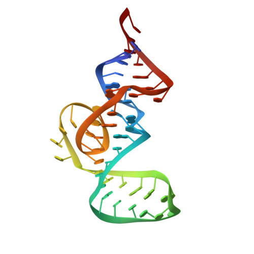
    Pepper fluorescent RNAs are a recently reported bright, stable and multicolor fluorogenic aptamer tag that enable imaging of diverse RNAs in live cells. To investigate the molecular basis of the superior properties of Pepper, we determined the structures of complexes of Pepper aptamer bound with its cognate HBC or HBC-like fluorophores at high resolution by X-ray crystallography. The Pepper aptamer folds in a monomeric non-G-quadruplex tuning-fork-like architecture composed of a helix and one protruded junction region. The near-planar fluorophore molecule intercalates in the middle of the structure and is sandwiched between one non-G-quadruplex base quadruple and one noncanonical G·U wobble helical base pair. In addition, structure-based mutational analysis is evaluated by in vitro and live-cell fluorogenic detection. Taken together, our research provides a structural basis for demystifying the fluorescence activation mechanism of Pepper aptamer and for further improvement of its future application in RNA visualization.


  • Organizational Affiliation
    • Life Sciences Institute, Zhejiang University, Hangzhou, China.

Macromolecule Content 

  • Total Structure Weight: 16.44 kDa 
  • Atom Count: 1,235 
  • Modeled Residue Count: 48 
  • Deposited Residue Count: 48 
  • Unique nucleic acid chains: 1

Macromolecules

Find similar nucleic acids by:  (by identity cutoff) 
Entity ID: 1
MoleculeChains LengthOrganismImage
Pepper (49-MER)48synthetic construct
Sequence Annotations
Expand
Reference Sequence

Small Molecules

Ligands 3 Unique
IDChains Name / Formula / InChI Key2D Diagram3D Interactions
GTP

Query on GTP


Download:Ideal Coordinates CCD File
B [auth A]GUANOSINE-5'-TRIPHOSPHATE
C10 H16 N5 O14 P3
XKMLYUALXHKNFT-UUOKFMHZSA-N
J93
(Subject of Investigation/LOI)

Query on J93


Download:Ideal Coordinates CCD File
H [auth A]4-[(~{Z})-1-cyano-2-[5-[2-hydroxyethyl(methyl)amino]thieno[3,2-b]thiophen-2-yl]ethenyl]benzenecarbonitrile
C19 H15 N3 O S2
RIGJCKIIBUZTCX-OVCLIPMQSA-N
MG
(Subject of Investigation/LOI)

Query on MG


Download:Ideal Coordinates CCD File
C [auth A],
D [auth A],
E [auth A],
F [auth A],
G [auth A]
MAGNESIUM ION
Mg
JLVVSXFLKOJNIY-UHFFFAOYSA-N

Experimental Data & Validation

Experimental Data

  • Method: X-RAY DIFFRACTION
  • Resolution: 1.80 Å
  • R-Value Free:  0.211 (Depositor), 0.212 (DCC) 
  • R-Value Work:  0.190 (Depositor), 0.191 (DCC) 
  • R-Value Observed: 0.191 (Depositor) 
Space Group: C 1 2 1
Unit Cell:
Length ( Å )Angle ( ˚ )
a = 92.447α = 90
b = 34.983β = 127.57
c = 57.839γ = 90
Software Package:
Software NamePurpose
HKL-2000data scaling
PHENIXrefinement
PDB_EXTRACTdata extraction
HKL-3000data reduction
PHASERphasing

Structure Validation

View Full Validation Report


Ligand Structure Quality Assessment 


Entry History 

& Funding Information

Deposition Data


Funding OrganizationLocationGrant Number
National Natural Science Foundation of China (NSFC)China--

Revision History  (Full details and data files)

  • Version 1.0: 2021-11-24
    Type: Initial release
  • Version 1.1: 2022-02-16
    Changes: Database references
  • Version 1.2: 2023-11-29
    Changes: Data collection, Refinement description