7EMJ

Crystal structure of T2R-TTL-Barbigerone complex


Experimental Data Snapshot

  • Method: X-RAY DIFFRACTION
  • Resolution: 2.33 Å
  • R-Value Free: 0.241 
  • R-Value Work: 0.198 
  • R-Value Observed: 0.200 

wwPDB Validation   3D Report Full Report


Ligand Structure Quality Assessment 


This is version 1.2 of the entry. See complete history


Literature

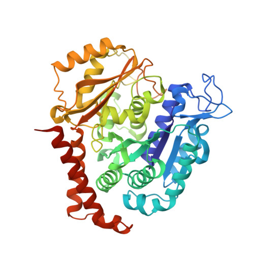
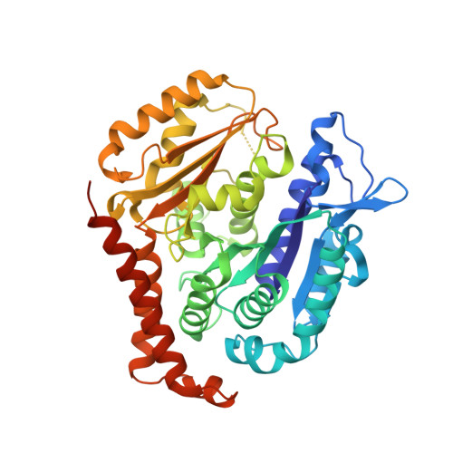
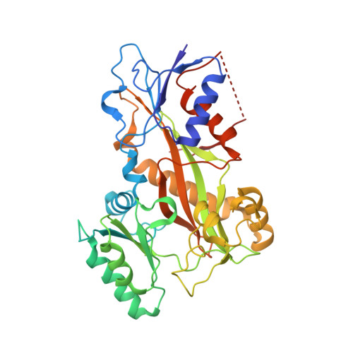
Crystal structure of tubulin-barbigerone complex enables rational design of potent anticancer agents with isoflavone skeleton.

Yan, W.Li, Y.Liu, Y.Wen, Y.Pei, H.Yang, J.Chen, L.

(2023) Phytomedicine 109: 154550-154550

  • DOI: https://doi.org/10.1016/j.phymed.2022.154550
  • Primary Citation of Related Structures:  
    7EMJ

  • PubMed Abstract: 

    Isoflavones possess many biological activities, including anti-inflammatory and anticancer effects. Microtubules (composed of αβ-tubulin heterodimers) are described as one possible cellular target of some of these isoflavones. However, the binding of tubulin to isoflavones has not been extensively studied, and until now, no crystal structure of the tubulin-isoflavone complex has been solved, and details of the isoflavone-tubulin interaction remain elusive. Barbigerone is an isoflavone mainly found in the genus Milletti, such as the edible leguminous plant Millettia ferruginea, with anticancer activity. This study aims to confirm the cellular target of barbigerone and to study its anticancer mechanism. Surface plasmon resonance assays and X-ray crystallography were used to study the interaction of barbigerone with tubulin protein. Immunofluorescence, in-cell and in vitro tubulin polymerization assays were employed to investigate the mechanism. MTT assays, cell clonal formation assays, wound healing assays, tube formation assays and H460 xenograft models were conducted to evaluate the in vitro and in vivo anticancer activities of barbigerone and one of its derivatives, 0412. Here, we found that barbigerone binds to tubulin to inhibit tubulin polymerization. Moreover, we solved the X-ray crystal structure of the tubulin-barbigerone complex at 2.33 Å resolution, which unambiguously determined the orientation and position of barbigerone in the colchicine-binding site. Illuminated by the X-ray data, we synthetized and obtained a more active isoflavone, 0412. Both barbigerone and 0412 inhibit cancer cell proliferation, tubulin polymerization, migration of HeLa cells and capillary-like tube formation of HUVECs, induce G2/M phase cell cycle arrest and apoptosis, and exhibit anticancer activity in an H460 xenograft model. In all, through biochemical and X-ray crystal structure results, we identified tubulin as the cellular target of one isoflavone, barbigerone, and proved that the tubulin-barbigerone complex plays a guiding role in obtaining a more active compound, 0412. These studies provide a crucial research basis for the development of isoflavones as anticancer candidate compounds.


  • Organizational Affiliation

    Laboratory of Natural and targeted small molecule drugs, State Key Laboratory of Biotherapy and Cancer Center, National Clinical Research Center for Geriatrics, West China Hospital, Sichuan University and Collaborative Innovation Center of Biotherapy, Chengdu 610041, China.


Macromolecules
Find similar proteins by:  (by identity cutoff)  |  3D Structure
Entity ID: 1
MoleculeChains Sequence LengthOrganismDetailsImage
Tubulin alpha-1B chain
A, C
451Sus scrofaMutation(s): 0 
UniProt
Find proteins for Q2XVP4 (Sus scrofa)
Explore Q2XVP4 
Go to UniProtKB:  Q2XVP4
Entity Groups  
Sequence Clusters30% Identity50% Identity70% Identity90% Identity95% Identity100% Identity
UniProt GroupQ2XVP4
Sequence Annotations
Expand
  • Reference Sequence
Find similar proteins by:  (by identity cutoff)  |  3D Structure
Entity ID: 2
MoleculeChains Sequence LengthOrganismDetailsImage
Tubulin beta chain
B, D
445Sus scrofaMutation(s): 0 
UniProt
Find proteins for P02554 (Sus scrofa)
Explore P02554 
Go to UniProtKB:  P02554
Entity Groups  
Sequence Clusters30% Identity50% Identity70% Identity90% Identity95% Identity100% Identity
UniProt GroupP02554
Sequence Annotations
Expand
  • Reference Sequence
Find similar proteins by:  (by identity cutoff)  |  3D Structure
Entity ID: 3
MoleculeChains Sequence LengthOrganismDetailsImage
Stathmin-4189Rattus norvegicusMutation(s): 0 
Gene Names: Stmn4
UniProt
Find proteins for P63043 (Rattus norvegicus)
Explore P63043 
Go to UniProtKB:  P63043
Entity Groups  
Sequence Clusters30% Identity50% Identity70% Identity90% Identity95% Identity100% Identity
UniProt GroupP63043
Sequence Annotations
Expand
  • Reference Sequence
Find similar proteins by:  (by identity cutoff)  |  3D Structure
Entity ID: 4
MoleculeChains Sequence LengthOrganismDetailsImage
Tubulin tyrosine ligase384Gallus gallusMutation(s): 0 
Gene Names: TTL
Entity Groups  
Sequence Clusters30% Identity50% Identity70% Identity90% Identity95% Identity100% Identity
Sequence Annotations
Expand
  • Reference Sequence
Small Molecules
Ligands 7 Unique
IDChains Name / Formula / InChI Key2D Diagram3D Interactions
GTP
Query on GTP

Download Ideal Coordinates CCD File 
G [auth A],
Q [auth C],
T [auth D]
GUANOSINE-5'-TRIPHOSPHATE
C10 H16 N5 O14 P3
XKMLYUALXHKNFT-UUOKFMHZSA-N
ACP
Query on ACP

Download Ideal Coordinates CCD File 
W [auth F]PHOSPHOMETHYLPHOSPHONIC ACID ADENYLATE ESTER
C11 H18 N5 O12 P3
UFZTZBNSLXELAL-IOSLPCCCSA-N
GDP
Query on GDP

Download Ideal Coordinates CCD File 
P [auth B]GUANOSINE-5'-DIPHOSPHATE
C10 H15 N5 O11 P2
QGWNDRXFNXRZMB-UUOKFMHZSA-N
J6L (Subject of Investigation/LOI)
Query on J6L

Download Ideal Coordinates CCD File 
O [auth B]8,8-dimethyl-3-(2,4,5-trimethoxyphenyl)pyrano[2,3-f]chromen-4-one
C23 H22 O6
OBIUGMGQVQMVSK-UHFFFAOYSA-N
MES
Query on MES

Download Ideal Coordinates CCD File 
L [auth B]2-(N-MORPHOLINO)-ETHANESULFONIC ACID
C6 H13 N O4 S
SXGZJKUKBWWHRA-UHFFFAOYSA-N
CA
Query on CA

Download Ideal Coordinates CCD File 
I [auth A]
J [auth A]
M [auth B]
N [auth B]
S [auth C]
I [auth A],
J [auth A],
M [auth B],
N [auth B],
S [auth C],
V [auth D]
CALCIUM ION
Ca
BHPQYMZQTOCNFJ-UHFFFAOYSA-N
MG
Query on MG

Download Ideal Coordinates CCD File 
H [auth A],
K [auth B],
R [auth C],
U [auth D],
X [auth F]
MAGNESIUM ION
Mg
JLVVSXFLKOJNIY-UHFFFAOYSA-N
Experimental Data & Validation

Experimental Data

  • Method: X-RAY DIFFRACTION
  • Resolution: 2.33 Å
  • R-Value Free: 0.241 
  • R-Value Work: 0.198 
  • R-Value Observed: 0.200 
  • Space Group: P 21 21 21
Unit Cell:
Length ( Å )Angle ( ˚ )
a = 105.03α = 90
b = 157.252β = 90
c = 180.606γ = 90
Software Package:
Software NamePurpose
PHENIXrefinement
HKL-3000data reduction
HKL-2000data scaling
HKL-3000phasing

Structure Validation

View Full Validation Report


Ligand Structure Quality Assessment 


Entry History 

Deposition Data

Revision History  (Full details and data files)

  • Version 1.0: 2022-04-20
    Type: Initial release
  • Version 1.1: 2023-11-29
    Changes: Data collection, Refinement description
  • Version 1.2: 2024-07-10
    Changes: Database references