7EJS | pdb_00007ejs

Structure of ERH-2 bound to PICS-1


Experimental Data Snapshot

  • Method: X-RAY DIFFRACTION
  • Resolution: 2.39 Å
  • R-Value Free: 
    0.238 (Depositor), 0.226 (DCC) 
  • R-Value Work: 
    0.201 (Depositor) 
  • R-Value Observed: 
    0.205 (Depositor) 

Starting Model: experimental
View more details

wwPDB Validation 3D Report Full Report

Validation slider image for 7EJS

This is version 1.2 of the entry. See complete history

Literature

Molecular basis for PICS-mediated piRNA biogenesis and cell division.

Wang, X.Zeng, C.Liao, S.Zhu, Z.Zhang, J.Tu, X.Yao, X.Feng, X.Guang, S.Xu, C.

(2021) Nat Commun 12: 5595-5595

  • DOI: https://doi.org/10.1038/s41467-021-25896-7
  • Primary Citation Related Structures: 
    7D1L, 7D2Y, 7EJO, 7EJS

  • PubMed Abstract: 

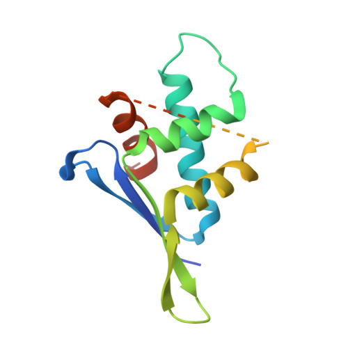
    By incorporating two mutually exclusive factors, PID-1 and TOST-1, C. elegans PICS complex plays important roles in piRNA biogenesis, chromosome segregation and cell division. We firstly map the interaction network between PICS subunits, then uncover the mechanisms underlying the interactions between PICS subunits by solving several complex structures, including those of TOFU-6/PICS-1, ERH-2/PICS-1, and ERH-2/TOST-1. Our biochemical experiment also demonstrates that PICS exists as an octamer consisting of two copies of each subunit. Combining structural analyses with mutagenesis experiments, we identify interfacial residues of PICS subunits that are critical for maintaining intact PICS complex in vitro. Furthermore, using genetics, cell biology and imaging experiments, we find that those mutants impairing the in vitro interaction network within PICS, also lead to dysfunction of PICS in vivo, including mislocalization of PICS, and reduced levels of piRNAs or aberrant chromosome segregation and cell division. Therefore, our work provides structural insights into understanding the PICS-mediated piRNA biogenesis and cell division.


  • Organizational Affiliation
    • Ministry of Education Key Laboratory for Membraneless Organelles & Cellular Dynamics, Hefei National Laboratory for Physical Sciences at the Microscale, School of Life Sciences, Division of Life Sciences and Medicine, University of Science and Technology of China, Hefei, PR China.

Macromolecule Content 

  • Total Structure Weight: 31.89 kDa 
  • Atom Count: 1,679 
  • Modeled Residue Count: 217 
  • Deposited Residue Count: 290 
  • Unique protein chains: 1

Macromolecules

Find similar proteins by:|  3D Structure
Entity ID: 1
MoleculeChains  Sequence LengthOrganismDetailsImage
Enhancer of rudimentary homolog 2,Protein pid-3
A, B
145Caenorhabditis elegansMutation(s): 0 
Gene Names: erh-2F35G12.11pid-3pics-1Y23H5A.3
UniProt
Find proteins for O76616 (Caenorhabditis elegans)
Explore O76616 
Go to UniProtKB:  O76616
Find proteins for Q20057 (Caenorhabditis elegans)
Explore Q20057 
Go to UniProtKB:  Q20057
Entity Groups
Sequence Clusters30% Identity50% Identity70% Identity90% Identity95% Identity100% Identity
UniProt GroupsO76616Q20057
Sequence Annotations
Expand
Reference Sequence

Experimental Data & Validation

Experimental Data

  • Method: X-RAY DIFFRACTION
  • Resolution: 2.39 Å
  • R-Value Free:  0.238 (Depositor), 0.226 (DCC) 
  • R-Value Work:  0.201 (Depositor) 
  • R-Value Observed: 0.205 (Depositor) 
Space Group: P 21 21 21
Unit Cell:
Length ( Å )Angle ( ˚ )
a = 44.825α = 90
b = 52.528β = 90
c = 125.119γ = 90
Software Package:
Software NamePurpose
PHENIXrefinement
PDB_EXTRACTdata extraction
HKL-3000data reduction
HKL-3000data scaling
PHASERphasing

Structure Validation

View Full Validation Report


Entry History 

Deposition Data

Revision History  (Full details and data files)

  • Version 1.0: 2021-08-25
    Type: Initial release
  • Version 1.1: 2021-10-06
    Changes: Database references
  • Version 1.2: 2023-11-29
    Changes: Data collection, Refinement description