7EDD | pdb_00007edd

Crystal structure of a serine protease from Streptococcus pyogenes


Experimental Data Snapshot

  • Method: X-RAY DIFFRACTION
  • Resolution: 2.90 Å
  • R-Value Free: 
    0.255 (Depositor), 0.252 (DCC) 
  • R-Value Work: 
    0.195 (Depositor), 0.199 (DCC) 
  • R-Value Observed: 
    0.198 (Depositor) 

Starting Model: experimental
View more details

wwPDB Validation 3D Report Full Report

Validation slider image for 7EDD

This is version 1.3 of the entry. See complete history

Literature

The Autocatalytic Cleavage Domain Is Not Required for the Activity of ScpC, a Virulence Protease from Streptococcus pyogenes : A Structural Insight.

Jobichen, C.Ying Chong, T.Hui Ling, T.Sivaraman, J.

(2021) Biochemistry 60: 1564-1568

  • DOI: https://doi.org/10.1021/acs.biochem.1c00185
  • Primary Citation Related Structures: 
    7EDD

  • PubMed Abstract: 

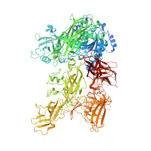
    Group A Streptococcus (GAS, or Streptococcus pyogenes ) is a leading human bacterial pathogen with diverse clinical manifestations, ranging from mild to life-threatening and to severe immune sequela. These diseases, combined, account for more than half a million deaths per year, globally. To accomplish its vast pathogenic potential, GAS expresses a multitude of virulent proteins, including the pivotal virulence factor ScpC. ScpC is a narrow-range surface-exposed subtilisin-like serine protease that cleaves the last 14 C-terminal amino acids of interleukin 8 (IL-8 or CXCL8) and impairs essential IL-8 signaling processes. As a result, neutrophil migration, bacterial killing, and the formation of neutrophil extracellular traps are strongly impaired. Also, ScpC has been identified as a potential vaccine candidate. ScpC undergoes an autocatalytic cleavage between Gln244 and Ser245, resulting in two polypeptide chains that assemble together forming the active protease. Previously, we reported that the region harboring the autocatalytic cleavage site, stretching from Gln213 to Asp272, is completely disordered. Here, we show that a deletion mutant (ScpC Δ60 ) of this region forms a single polypeptide chain, whose crystal structure we determined at 2.9 Å resolution. Moreover, we show that ScpC Δ60 is an active protease capable of cleaving its substrate IL-8 in a manner comparable to that of the wild type. These studies improve our understanding of the proteolytic activity of ScpC.


  • Organizational Affiliation
    • Department of Biological Sciences, National University of Singapore, 14 Science Drive 4, Singapore 117543.

Macromolecule Content 

  • Total Structure Weight: 169.56 kDa 
  • Atom Count: 10,868 
  • Modeled Residue Count: 1,394 
  • Deposited Residue Count: 1,519 
  • Unique protein chains: 1

Macromolecules

Find similar proteins by:|  3D Structure
Entity ID: 1
MoleculeChains  Sequence LengthOrganismDetailsImage
C5a peptidase1,519Streptococcus pyogenesMutation(s): 0 
Gene Names: scpC
EC: 3.4.21.110
UniProt
Find proteins for Q3HV58 (Streptococcus pyogenes)
Explore Q3HV58 
Go to UniProtKB:  Q3HV58
Entity Groups
Sequence Clusters30% Identity50% Identity70% Identity90% Identity95% Identity100% Identity
UniProt GroupQ3HV58
Sequence Annotations
Expand
Reference Sequence

Small Molecules

Ligands 6 Unique
IDChains Name / Formula / InChI Key2D Diagram3D Interactions
PEG

Query on PEG


Download:Ideal Coordinates CCD File
T [auth A]DI(HYDROXYETHYL)ETHER
C4 H10 O3
MTHSVFCYNBDYFN-UHFFFAOYSA-N
SO4

Query on SO4


Download:Ideal Coordinates CCD File
B [auth A],
C [auth A],
D [auth A]
SULFATE ION
O4 S
QAOWNCQODCNURD-UHFFFAOYSA-L
GOL

Query on GOL


Download:Ideal Coordinates CCD File
N [auth A],
O [auth A],
P [auth A],
Q [auth A],
R [auth A]
GLYCEROL
C3 H8 O3
PEDCQBHIVMGVHV-UHFFFAOYSA-N
CA

Query on CA


Download:Ideal Coordinates CCD File
E [auth A]
F [auth A]
G [auth A]
H [auth A]
I [auth A]
E [auth A],
F [auth A],
G [auth A],
H [auth A],
I [auth A],
J [auth A],
K [auth A],
L [auth A],
M [auth A],
W [auth A]
CALCIUM ION
Ca
BHPQYMZQTOCNFJ-UHFFFAOYSA-N
CL

Query on CL


Download:Ideal Coordinates CCD File
U [auth A],
V [auth A]
CHLORIDE ION
Cl
VEXZGXHMUGYJMC-UHFFFAOYSA-M
MG

Query on MG


Download:Ideal Coordinates CCD File
S [auth A]MAGNESIUM ION
Mg
JLVVSXFLKOJNIY-UHFFFAOYSA-N
Modified Residues  1 Unique
IDChains TypeFormula2D DiagramParent
MSE
Query on MSE
A
L-PEPTIDE LINKINGC5 H11 N O2 SeMET

Experimental Data & Validation

Experimental Data

  • Method: X-RAY DIFFRACTION
  • Resolution: 2.90 Å
  • R-Value Free:  0.255 (Depositor), 0.252 (DCC) 
  • R-Value Work:  0.195 (Depositor), 0.199 (DCC) 
  • R-Value Observed: 0.198 (Depositor) 
Space Group: P 62 2 2
Unit Cell:
Length ( Å )Angle ( ˚ )
a = 191.969α = 90
b = 191.969β = 90
c = 252.788γ = 120
Software Package:
Software NamePurpose
PHENIXrefinement
HKL-2000data reduction
HKL-2000data scaling
PHENIXphasing

Structure Validation

View Full Validation Report


Entry History 

Deposition Data

Revision History  (Full details and data files)

  • Version 1.0: 2021-05-12
    Type: Initial release
  • Version 1.1: 2021-05-26
    Changes: Database references
  • Version 1.2: 2023-11-29
    Changes: Data collection, Database references, Refinement description
  • Version 1.3: 2024-11-20
    Changes: Structure summary