7ED6 | pdb_00007ed6

Crystal structure of Thermus thermophilus FakA ATP-binding domain


Experimental Data Snapshot

  • Method: X-RAY DIFFRACTION
  • Resolution: 1.93 Å
  • R-Value Free: 
    0.236 (Depositor), 0.243 (DCC) 
  • R-Value Work: 
    0.208 (Depositor), 0.212 (DCC) 
  • R-Value Observed: 
    0.209 (Depositor) 

wwPDB Validation 3D Report Full Report

Validation slider image for 7ED6

Ligand Structure Quality Assessment 


This is version 1.2 of the entry. See complete history

Literature

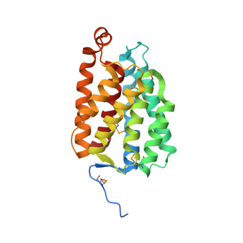
Crystal structure of a nucleotide-binding domain of fatty acid kinase FakA from Thermus thermophilus HB8.

Nakatani, M.Nakahara, S.Y.Fukui, K.Urano, M.Fujii, Y.Murakawa, T.Baba, S.Kumasaka, T.Okanishi, H.Kanai, Y.Yano, T.Masui, R.

(2022) J Struct Biol 214: 107904-107904

  • DOI: https://doi.org/10.1016/j.jsb.2022.107904
  • Primary Citation Related Structures: 
    7ED6, 7ED9

  • PubMed Abstract: 

    Fatty acid kinase is necessary for the incorporation of exogenous fatty acids into membrane phospholipids. Fatty acid kinase consists of two components: a kinase component, FakA, that phosphorylates a fatty acid bound to a fatty acid-binding component, FakB. However, the molecular details underlying the phosphotransfer reaction remain to be resolved. We determined the crystal structure of the N-terminal domain of FakA bound to ADP from Thermus thermophilus HB8. The overall structure of this domain showed that the helical barrel fold is similar to the nucleotide-binding component of dihydroxyacetone kinase. The structure of the nucleotide-binding site revealed the roles of the conserved residues in recognition of ADP and Mg 2+ , but the N-terminal domain of FakA lacked the ADP-capping loop found in the dihydroxyacetone kinase component. Based on the structural similarity to the two subunits of dihydroxyacetone kinase complex, we constructed a model of the complex of T. thermophilus FakB and the N-terminal domain of FakA. In this model, the invariant Arg residue of FakB occupied a position that was spatially similar to that of the catalytically important Arg residue of dihydroxyacetone kinase, which predicted a composite active site in the Fatty acid kinase complex.


  • Organizational Affiliation
    • Graduate School of Science, Osaka City University, 3-3-138 Sugimoto, Sumiyoshi-ku, Osaka 558-8585, Japan.

Macromolecule Content 

  • Total Structure Weight: 49 kDa 
  • Atom Count: 3,144 
  • Modeled Residue Count: 399 
  • Deposited Residue Count: 438 
  • Unique protein chains: 1

Macromolecules

Find similar proteins by:|  3D Structure
Entity ID: 1
MoleculeChains  Sequence LengthOrganismDetailsImage
Probable kinase
A, B
219Thermus thermophilus HB8Mutation(s): 0 
Gene Names: TTHA0214
UniProt
Find proteins for Q5SLS9 (Thermus thermophilus (strain ATCC 27634 / DSM 579 / HB8))
Explore Q5SLS9 
Go to UniProtKB:  Q5SLS9
Entity Groups
Sequence Clusters30% Identity50% Identity70% Identity90% Identity95% Identity100% Identity
UniProt GroupQ5SLS9
Sequence Annotations
Expand
Reference Sequence

Experimental Data & Validation

Experimental Data

  • Method: X-RAY DIFFRACTION
  • Resolution: 1.93 Å
  • R-Value Free:  0.236 (Depositor), 0.243 (DCC) 
  • R-Value Work:  0.208 (Depositor), 0.212 (DCC) 
  • R-Value Observed: 0.209 (Depositor) 
Space Group: P 43 21 2
Unit Cell:
Length ( Å )Angle ( ˚ )
a = 90.19α = 90
b = 90.19β = 90
c = 111.62γ = 90
Software Package:
Software NamePurpose
PHENIXrefinement
XDSdata processing
XDSdata scaling
PHENIXphasing
PHENIXmodel building
Cootmodel building
XDSdata reduction

Structure Validation

View Full Validation Report


Ligand Structure Quality Assessment 


Entry History 

Deposition Data

Revision History  (Full details and data files)

  • Version 1.0: 2022-03-16
    Type: Initial release
  • Version 1.1: 2023-01-11
    Changes: Database references
  • Version 1.2: 2024-11-06
    Changes: Data collection, Refinement description, Structure summary