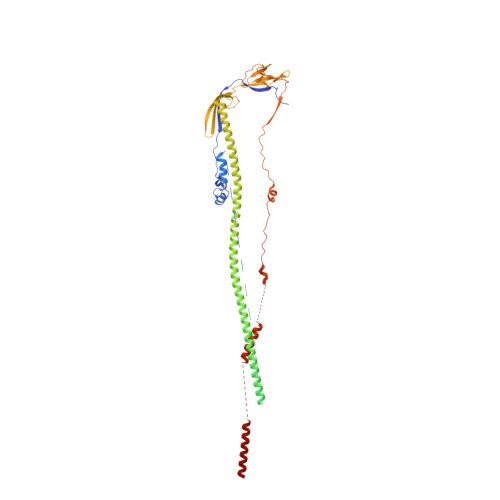
7E9T | pdb_00007e9t

Nanometer resolution in situ structure of SARS-CoV-2 post-fusion spike


Experimental Data Snapshot

  • Method: ELECTRON MICROSCOPY
  • Resolution: 10.9 Å
  • Aggregation State: PARTICLE 
  • Reconstruction Method: SUBTOMOGRAM AVERAGING 

wwPDB Validation 3D Report Full Report

Validation slider image for 7E9T

This is version 1.3 of the entry. See complete history. 

Literature

Nanometer-resolution in situ structure of the SARS-CoV-2 postfusion spike protein.

Tai, L., Zhu, G., Yang, M., Cao, L., Xing, X., Yin, G., Chan, C., Qin, C., Rao, Z., Wang, X., Sun, F., Zhu, Y.

(2021) Proc Natl Acad Sci U S A 118

  • DOI: https://doi.org/10.1073/pnas.2112703118
  • Primary Citation Related Structures: 
    7E9T

  • PubMed Abstract: 

    The spike protein of severe acute respiratory syndrome coronavirus 2 (SARS-CoV-2) mediates membrane fusion to allow entry of the viral genome into host cells. To understand its detailed entry mechanism and develop a specific entry inhibitor, in situ structural information on the SARS-CoV-2 spike protein in different states is urgent. Here, by using cryo-electron tomography, we observed both prefusion and postfusion spikes in β-propiolactone-inactivated SARS-CoV-2 virions and solved the in situ structure of the postfusion spike at nanometer resolution. Compared to previous reports, the six-helix bundle fusion core, the glycosylation sites, and the location of the transmembrane domain were clearly resolved. We observed oligomerization patterns of the spikes on the viral membrane, likely suggesting a mechanism of fusion pore formation.


  • Organizational Affiliation: 
    • National Key Laboratory of Biomacromolecules, CAS Center for Excellence in Biomacromolecules, Institute of Biophysics, Chinese Academy of Sciences, Beijing 100101, China.

Macromolecule Content 

  • Total Structure Weight: 184.56 kDa 
  • Atom Count: 9,198 
  • Modeled Residue Count: 1,113 
  • Deposited Residue Count: 1,596 
  • Unique protein chains: 1

Macromolecules

Find similar proteins by:|  3D Structure
Entity ID: 1
MoleculeChains  Sequence LengthOrganismDetailsImage
Spike protein S2
A, B, C
532Severe acute respiratory syndrome coronavirus 2Mutation(s): 0 
Membrane Entity: Yes 
UniProt
Find proteins for P0DTC2 (Severe acute respiratory syndrome coronavirus 2)
Explore P0DTC2 
Go to UniProtKB:  P0DTC2
Entity Groups
Sequence Clusters30% Identity50% Identity70% Identity90% Identity95% Identity100% Identity
UniProt GroupP0DTC2
Glycosylation
Glycosylation Sites: 6Go to GlyGen: P0DTC2-1
Sequence Annotations
Expand
Reference Sequence

Oligosaccharides

Help  
Entity ID: 2
MoleculeChains Length2D Diagram GlycosylationD Interactions
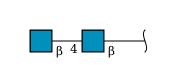
2-acetamido-2-deoxy-beta-D-glucopyranose-(1-4)-2-acetamido-2-deoxy-beta-D-glucopyranoseD,
G [auth H],
J [auth L]
2N-Glycosylation
Glycosylation Resources
GlyTouCan: G42666HT
GlyCosmos: G42666HT
GlyGen: G42666HT
Entity ID: 3
MoleculeChains Length2D Diagram GlycosylationD Interactions
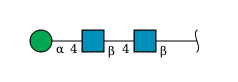
alpha-D-mannopyranose-(1-4)-2-acetamido-2-deoxy-beta-D-glucopyranose-(1-4)-2-acetamido-2-deoxy-beta-D-glucopyranose
E,
F,
H [auth I],
I [auth J],
K [auth M],
E,
F,
H [auth I],
I [auth J],
K [auth M],
L [auth N],
M [auth K],
N [auth O],
O [auth G]
3N-Glycosylation
Glycosylation Resources
GlyTouCan: G62182OO
GlyCosmos: G62182OO
GlyGen: G62182OO

Small Molecules

Ligands 1 Unique
IDChains Name / Formula / InChI Key2D Diagram3D Interactions
NAG

Query on NAG


Download:Ideal Coordinates CCD File
P [auth A]
Q [auth A]
R [auth A]
S [auth B]
T [auth B]
P [auth A],
Q [auth A],
R [auth A],
S [auth B],
T [auth B],
U [auth B],
V [auth C],
W [auth C],
X [auth C]
2-acetamido-2-deoxy-beta-D-glucopyranose
C8 H15 N O6
OVRNDRQMDRJTHS-FMDGEEDCSA-N

Experimental Data & Validation

Experimental Data

  • Method: ELECTRON MICROSCOPY
  • Resolution: 10.9 Å
  • Aggregation State: PARTICLE 
  • Reconstruction Method: SUBTOMOGRAM AVERAGING 
EM Software:
TaskSoftware PackageVersion
MODEL REFINEMENTPHENIX

Structure Validation

View Full Validation Report


Entry History 

Deposition Data

Revision History  (Full details and data files)

  • Version 1.0: 2021-11-17
    Type: Initial release
  • Version 1.1: 2022-03-23
    Changes: Database references
  • Version 1.2: 2024-10-16
    Changes: Data collection, Structure summary
  • Version 1.3: 2025-06-18
    Changes: Data collection