7E62 | pdb_00007e62

Mouse TAB2 NZF in complex with Lys6-linked diubiquitin


Experimental Data Snapshot

  • Method: X-RAY DIFFRACTION
  • Resolution: 1.99 Å
  • R-Value Free: 
    0.264 (Depositor), 0.265 (DCC) 
  • R-Value Work: 
    0.242 (Depositor), 0.240 (DCC) 
  • R-Value Observed: 
    0.243 (Depositor) 

Starting Model: experimental
View more details

wwPDB Validation 3D Report Full Report

Validation slider image for 7E62

This is version 1.2 of the entry. See complete history

Literature

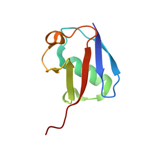
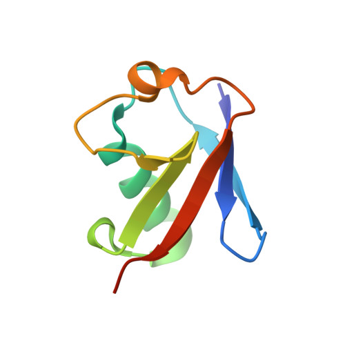
Structural basis for specific recognition of K6-linked polyubiquitin chains by the TAB2 NZF domain.

Li, Y.Okatsu, K.Fukai, S.Sato, Y.

(2021) Biophys J 120: 3355-3362

  • DOI: https://doi.org/10.1016/j.bpj.2021.06.037
  • Primary Citation Related Structures: 
    7E62

  • PubMed Abstract: 

    TAK1-binding protein 2 (TAB2) has generally been considered to bind specifically to K63-linked polyubiquitin chains via its C-terminal Npl4 zinc-finger (NZF) domain. However, a recent study showed that the NZF domain of TAB2 (TAB2-NZF) could also interact with K6-linked polyubiquitin chains. Here, we report the crystal structure of TAB2-NZF in complex with K6-linked diubiquitin (K6-Ub 2 ) at 1.99-Å resolution. TAB2-NZF simultaneously interacts with the distal and proximal ubiquitin moieties of K6-Ub 2 . By comparing the structures of TAB2-NZF in complex with K6-Ub 2 and with K63-linked diubiquitin (K63-Ub 2 ), we reveal that the binding mechanism of TAB2-NZF with K6-Ub 2 is similar to that with K63-Ub 2 , except for the flexible C-terminal region of the distal ubiquitin. Therefore, we conclude that the C-terminal flexibility of the distal ubiquitin contributes to the dual specificity of TAB2-NZF toward K6- and K63-linked ubiquitin chains. This study provides important insights into the functions of K6-linked ubiquitin chains, which are currently unclear.


  • Organizational Affiliation
    • Department of Computational Biology and Medical Sciences, Graduate School of Frontier Sciences, The University of Tokyo, Chiba, Japan.

Macromolecule Content 

  • Total Structure Weight: 47.35 kDa 
  • Atom Count: 2,932 
  • Modeled Residue Count: 359 
  • Deposited Residue Count: 416 
  • Unique protein chains: 3

Macromolecules

Find similar proteins by:|  3D Structure
Entity ID: 1
MoleculeChains  Sequence LengthOrganismDetailsImage
ubiquitinA,
D [auth H]
76Mus musculusMutation(s): 1 
Gene Names: Rps27aUba80Ubcep1
UniProt
Find proteins for P62983 (Mus musculus)
Explore P62983 
Go to UniProtKB:  P62983
Entity Groups
Sequence Clusters30% Identity50% Identity70% Identity90% Identity95% Identity100% Identity
UniProt GroupP62983
Sequence Annotations
Expand
Reference Sequence
Find similar proteins by:|  3D Structure
Entity ID: 2
MoleculeChains  Sequence LengthOrganismDetailsImage
UbiquitinB,
E [auth I]
77Mus musculusMutation(s): 1 
Gene Names: Rps27aUba80Ubcep1
UniProt
Find proteins for P62983 (Mus musculus)
Explore P62983 
Go to UniProtKB:  P62983
Entity Groups
Sequence Clusters30% Identity50% Identity70% Identity90% Identity95% Identity100% Identity
UniProt GroupP62983
Sequence Annotations
Expand
Reference Sequence
Find similar proteins by:|  3D Structure
Entity ID: 3
MoleculeChains  Sequence LengthOrganismDetailsImage
TGF-beta-activated kinase 1 and MAP3K7-binding protein 2C,
F [auth J]
55Mus musculusMutation(s): 0 
Gene Names: Tab2Kiaa0733Map3k7ip2
UniProt
Find proteins for Q99K90 (Mus musculus)
Explore Q99K90 
Go to UniProtKB:  Q99K90
Entity Groups
Sequence Clusters30% Identity50% Identity70% Identity90% Identity95% Identity100% Identity
UniProt GroupQ99K90
Sequence Annotations
Expand
Reference Sequence

Experimental Data & Validation

Experimental Data

  • Method: X-RAY DIFFRACTION
  • Resolution: 1.99 Å
  • R-Value Free:  0.264 (Depositor), 0.265 (DCC) 
  • R-Value Work:  0.242 (Depositor), 0.240 (DCC) 
  • R-Value Observed: 0.243 (Depositor) 
Space Group: P 21 21 21
Unit Cell:
Length ( Å )Angle ( ˚ )
a = 45.694α = 90
b = 76.17β = 90
c = 114.813γ = 90
Software Package:
Software NamePurpose
PHENIXrefinement
XDSdata reduction
XDSdata scaling
MOLREPphasing

Structure Validation

View Full Validation Report


Entry History 

& Funding Information

Deposition Data


Funding OrganizationLocationGrant Number
Japan Society for the Promotion of Science (JSPS)Japan18H05501

Revision History  (Full details and data files)

  • Version 1.0: 2021-08-18
    Type: Initial release
  • Version 1.1: 2021-11-10
    Changes: Database references
  • Version 1.2: 2023-11-29
    Changes: Data collection, Refinement description