


Structure of human RORgammat LBD with SRC2.2 at 2.80 Angstroms resolution
Liu, Z.H., Huang, J., Wu, Z.R., Tang, Y., Lu, W.Q.To be published.
Experimental Data Snapshot
Starting Model: experimental
View more details
Entity ID: 1 | |||||
|---|---|---|---|---|---|
| Molecule | Chains | Sequence Length | Organism | Details | Image |
| Nuclear receptor ROR-gamma | 243 | Homo sapiens | Mutation(s): 0 Gene Names: RORC, NR1F3, RORG, RZRG | 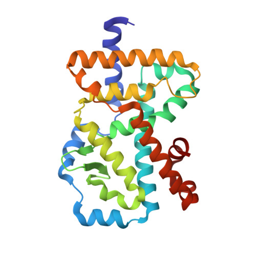 | |
UniProt & NIH Common Fund Data Resources | |||||
PHAROS: P51449 GTEx: ENSG00000143365 | |||||
Entity Groups | |||||
| Sequence Clusters | 30% Identity50% Identity70% Identity90% Identity95% Identity100% Identity | ||||
| UniProt Group | P51449 | ||||
Sequence AnnotationsExpand | |||||
Reference Sequence | |||||
Entity ID: 2 | |||||
|---|---|---|---|---|---|
| Molecule | Chains | Sequence Length | Organism | Details | Image |
| LYS-ILE-LEU-HIS-ARG-LEU-LEU-GLN | 8 | synthetic construct | Mutation(s): 0 | 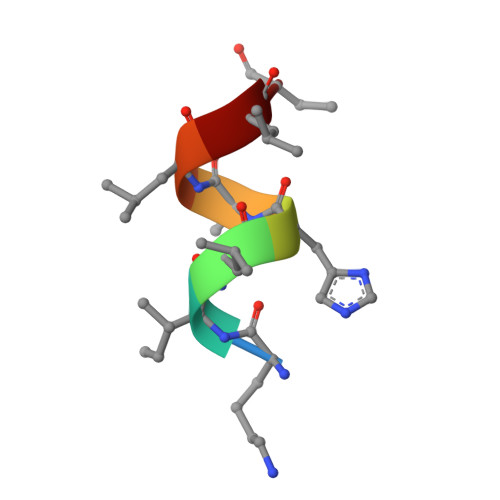 | |
| Ligands 1 Unique | |||||
|---|---|---|---|---|---|
| ID | Chains | Name / Formula / InChI Key | 2D Diagram | 3D Interactions | |
| HVL (Subject of Investigation/LOI) Download:Ideal Coordinates CCD File | C [auth A] | (3R,5R,6S,8R,9R,10R,12R,13R,14R,17S)-4,4,8,10,14-pentamethyl-17-[(2R)-2,6,6-trimethyloxan-2-yl]-2,3,5,6,7,9,11,12,13,15,16,17-dodecahydro-1H-cyclopenta[a]phenanthrene-3,6,12-triol C30 H52 O4 QFJUYMMIBFBOJY-FAKBLDBGSA-N |  | ||
| Length ( Å ) | Angle ( ˚ ) |
|---|---|
| a = 62.446 | α = 90 |
| b = 62.446 | β = 90 |
| c = 159.766 | γ = 90 |
| Software Name | Purpose |
|---|---|
| HKL-2000 | data reduction |
| HKL-2000 | data scaling |
| PHASER | phasing |
| REFMAC | refinement |
| PDB_EXTRACT | data extraction |


