7E3F | pdb_00007e3f

Crystal structure of Trypanosoma brucei cathepsin B Y217C/S275C mutant


Experimental Data Snapshot

  • Method: X-RAY DIFFRACTION
  • Resolution: 2.35 Å
  • R-Value Free: 
    0.257 (Depositor), 0.267 (DCC) 
  • R-Value Work: 
    0.219 (Depositor), 0.228 (DCC) 

Starting Model: experimental
View more details

wwPDB Validation 3D Report Full Report

Validation slider image for 7E3F

This is version 1.2 of the entry. See complete history

Literature

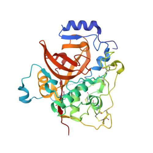
Design of an In-Cell Protein Crystal for the Environmentally Responsive Construction of a Supramolecular Filament.

Abe, S.Pham, T.T.Negishi, H.Yamashita, K.Hirata, K.Ueno, T.

(2021) Angew Chem Int Ed Engl 60: 12341-12345

  • DOI: https://doi.org/10.1002/anie.202102039
  • Primary Citation Related Structures: 
    7E3E, 7E3F, 7E3G

  • PubMed Abstract: 

    Protein assemblies can be designed for development of nano-bio materials. This has been achieved by modulating protein-protein interactions. However, fabrication of highly ordered protein assemblies remains challenging. Protein crystals, which have highly ordered arrangements of protein molecules, provide useful source matrices for synthesizing artificial protein assemblies. Here, we describe construction of a supramolecular filament structure by engineering covalent and non-covalent interactions in a protein crystal. Performing in-cell crystallization of Trypanosoma brucei cysteine protease cathepsin B (TbCatB), we achieved a precise arrangement of protein molecules while suppressing random aggregation due to disulfide bonds. We succeeded in synthesizing bundled filament from the crystals by autoxidation of cysteinyl thiols after the isolation of the crystals from living cells.


  • Organizational Affiliation
    • School of Life Science and Technology, Tokyo Institute of Technology, Nagatsuta 4259-B-55, Midori-ku, Yokohama, 226-8501, Japan.

Macromolecule Content 

  • Total Structure Weight: 35.39 kDa 
  • Atom Count: 2,482 
  • Modeled Residue Count: 308 
  • Deposited Residue Count: 312 
  • Unique protein chains: 1

Macromolecules

Find similar proteins by:|  3D Structure
Entity ID: 1
MoleculeChains  Sequence LengthOrganismDetailsImage
Cysteine peptidase C (CPC)312Trypanosoma brucei brucei TREU927Mutation(s): 2 
Gene Names: Tb927.6.560
EC: 3.4.22
UniProt
Find proteins for D6XHE1 (Trypanosoma brucei brucei (strain 927/4 GUTat10.1))
Explore D6XHE1 
Go to UniProtKB:  D6XHE1
Entity Groups
Sequence Clusters30% Identity50% Identity70% Identity90% Identity95% Identity100% Identity
UniProt GroupD6XHE1
Glycosylation
Glycosylation Sites: 2
Sequence Annotations
Expand
Reference Sequence

Oligosaccharides

Help  
Entity ID: 2
MoleculeChains Length2D Diagram GlycosylationD Interactions
2-acetamido-2-deoxy-beta-D-glucopyranose-(1-4)-2-acetamido-2-deoxy-beta-D-glucopyranose
B
2N-Glycosylation
Glycosylation Resources
GlyTouCan: G42666HT
GlyCosmos: G42666HT
GlyGen: G42666HT
Entity ID: 3
MoleculeChains Length2D Diagram GlycosylationD Interactions
beta-D-mannopyranose-(1-4)-2-acetamido-2-deoxy-beta-D-glucopyranose-(1-4)-2-acetamido-2-deoxy-beta-D-glucopyranose
C
3N-Glycosylation
Glycosylation Resources
GlyTouCan: G15407YE
GlyCosmos: G15407YE
GlyGen: G15407YE

Experimental Data & Validation

Experimental Data

  • Method: X-RAY DIFFRACTION
  • Resolution: 2.35 Å
  • R-Value Free:  0.257 (Depositor), 0.267 (DCC) 
  • R-Value Work:  0.219 (Depositor), 0.228 (DCC) 
Space Group: P 42 21 2
Unit Cell:
Length ( Å )Angle ( ˚ )
a = 123.65α = 90
b = 123.65β = 90
c = 54.28γ = 90
Software Package:
Software NamePurpose
REFMACrefinement
XSCALEdata scaling
PHENIXphasing
XDSdata processing

Structure Validation

View Full Validation Report


Entry History 

& Funding Information

Deposition Data


Funding OrganizationLocationGrant Number
Japan Society for the Promotion of Science (JSPS)JapanJP18H05421
Japan Society for the Promotion of Science (JSPS)JapanJP18K05140

Revision History  (Full details and data files)

  • Version 1.0: 2021-12-15
    Type: Initial release
  • Version 1.1: 2023-11-29
    Changes: Data collection, Derived calculations, Refinement description
  • Version 1.2: 2024-11-13
    Changes: Structure summary