


Crystal structure of a glutathione S-transferase mutant SbGSTU6(I55T) from Salix babylonica in complex with glutathione
Zhuge, X.L., Yang, H.L.To be published.
Experimental Data Snapshot
Starting Model: experimental
View more details
Entity ID: 1 | |||||
|---|---|---|---|---|---|
| Molecule | Chains | Sequence Length | Organism | Details | Image |
| Glutathione S-transferase | 235 | Salix babylonica | Mutation(s): 1 Gene Names: GSTU6 EC: 2.5.1.18 | 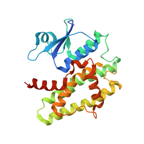 | |
UniProt | |||||
Entity Groups | |||||
| Sequence Clusters | 30% Identity50% Identity70% Identity90% Identity95% Identity100% Identity | ||||
| UniProt Group | A0A4Y5R032 | ||||
Sequence AnnotationsExpand | |||||
Reference Sequence | |||||
| Ligands 1 Unique | |||||
|---|---|---|---|---|---|
| ID | Chains | Name / Formula / InChI Key | 2D Diagram | 3D Interactions | |
| GSH (Subject of Investigation/LOI) Download:Ideal Coordinates CCD File | B [auth A] | Glutathione C10 H17 N3 O6 S RWSXRVCMGQZWBV-WDSKDSINSA-N |  | ||
| Entity ID: 2 | |||||
|---|---|---|---|---|---|
| ID | Chains | Name | Type/Class | 2D Diagram | 3D Interactions |
| PRD_002593 (GSH) Query on PRD_002593 | B [auth A] | Glutathione | Peptide-like / Oxidation-reduction | | |
| Length ( Å ) | Angle ( ˚ ) |
|---|---|
| a = 104.379 | α = 90 |
| b = 104.379 | β = 90 |
| c = 53.126 | γ = 120 |
| Software Name | Purpose |
|---|---|
| PHENIX | refinement |
| HKL-2000 | data reduction |
| HKL-2000 | data scaling |
| PHASER | phasing |
| Funding Organization | Location | Grant Number |
|---|---|---|
| National Natural Science Foundation of China (NSFC) | China | NSFC 32071486 |


