7DLK | pdb_00007dlk

Crystal Structure of veratryl alcohol bound Dye Decolorizing peroxidase from Bacillus subtilis


Experimental Data Snapshot

  • Method: X-RAY DIFFRACTION
  • Resolution: 2.10 Å
  • R-Value Free: 
    0.249 (Depositor), 0.248 (DCC) 
  • R-Value Work: 
    0.185 (Depositor), 0.186 (DCC) 
  • R-Value Observed: 
    0.188 (Depositor) 

Starting Model: experimental
View more details

wwPDB Validation 3D Report Full Report

Validation slider image for 7DLK

Ligand Structure Quality Assessment 


This is version 1.2 of the entry. See complete history

Literature

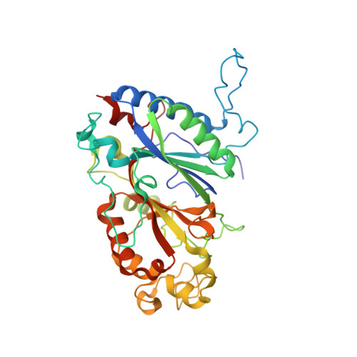
Structure of dye-decolorizing peroxidase from Bacillus subtilis in complex with veratryl alcohol.

Dhankhar, P.Dalal, V.Singh, V.Sharma, A.K.Kumar, P.

(2021) Int J Biol Macromol 193: 601-608

  • DOI: https://doi.org/10.1016/j.ijbiomac.2021.10.100
  • Primary Citation Related Structures: 
    7DLK

  • PubMed Abstract: 

    Dye-decolorizing peroxidases (DyPs) are heme-containing peroxidases, which have promising application in biodegradation of phenolic lignin compounds and in detoxification of dyes. In this study, the crystal structure of BsDyP- veratryl alcohol (VA) complex delves deep into the binding of small substrate molecules within the DyP heme cavity. The biochemical analysis shows that BsDyP oxidizes the VA with a turnover number of 0.065 s -1 , followed by the oxidation of 2,6-dimethoxyphenol (DMP) and guaiacol with a comparable turnover number (k cat ) of 0.07 s -1 and 0.07 s -1 , respectively. Moreover, biophysical and computational studies reveal the comparable binding affinity of substrates to BsDyP and produce lower-energy stable BsDyP-ligand(s) complexes. All together with our previous findings, we are providing a complete structural description of substrate-binding sites in DyP. The structural insight of BsDyP helps to modulate its engineering to enhance the activity towards the oxidation of a wide range of substrates.


  • Organizational Affiliation
    • Department of Biosciences and Bioengineering, Indian Institute of Technology Roorkee, 247667, India.

Macromolecule Content 

  • Total Structure Weight: 247.69 kDa 
  • Atom Count: 19,220 
  • Modeled Residue Count: 2,170 
  • Deposited Residue Count: 2,178 
  • Unique protein chains: 1

Macromolecules

Find similar proteins by:|  3D Structure
Entity ID: 1
MoleculeChains  Sequence LengthOrganismDetailsImage
Deferrochelatase/peroxidase
A, B, C, D, E
A, B, C, D, E, F
363Bacillus subtilisMutation(s): 0 
Gene Names: efeBGS595_16230
EC: 1.11.1 (PDB Primary Data), 4.98.1.1 (UniProt)
UniProt
Find proteins for P39597 (Bacillus subtilis (strain 168))
Explore P39597 
Go to UniProtKB:  P39597
Entity Groups
Sequence Clusters30% Identity50% Identity70% Identity90% Identity95% Identity100% Identity
UniProt GroupP39597
Sequence Annotations
Expand
Reference Sequence

Small Molecules

Ligands 8 Unique
IDChains Name / Formula / InChI Key2D Diagram3D Interactions
HEM
(Subject of Investigation/LOI)

Query on HEM


Download:Ideal Coordinates CCD File
EA [auth E]
G [auth A]
KA [auth F]
O [auth B]
S [auth C]
EA [auth E],
G [auth A],
KA [auth F],
O [auth B],
S [auth C],
Z [auth D]
PROTOPORPHYRIN IX CONTAINING FE
C34 H32 Fe N4 O4
KABFMIBPWCXCRK-RGGAHWMASA-L
EPE
(Subject of Investigation/LOI)

Query on EPE


Download:Ideal Coordinates CCD File
MA [auth F]4-(2-HYDROXYETHYL)-1-PIPERAZINE ETHANESULFONIC ACID
C8 H18 N2 O4 S
JKMHFZQWWAIEOD-UHFFFAOYSA-N
VOH
(Subject of Investigation/LOI)

Query on VOH


Download:Ideal Coordinates CCD File
GA [auth E],
U [auth C]
Veratryl alcohol
C9 H12 O3
OEGPRYNGFWGMMV-UHFFFAOYSA-N
MPD
(Subject of Investigation/LOI)

Query on MPD


Download:Ideal Coordinates CCD File
I [auth A],
V [auth C]
(4S)-2-METHYL-2,4-PENTANEDIOL
C6 H14 O2
SVTBMSDMJJWYQN-YFKPBYRVSA-N
PO4
(Subject of Investigation/LOI)

Query on PO4


Download:Ideal Coordinates CCD File
N [auth A]PHOSPHATE ION
O4 P
NBIIXXVUZAFLBC-UHFFFAOYSA-K
GOL
(Subject of Investigation/LOI)

Query on GOL


Download:Ideal Coordinates CCD File
BA [auth D]
HA [auth E]
IA [auth E]
J [auth A]
K [auth A]
BA [auth D],
HA [auth E],
IA [auth E],
J [auth A],
K [auth A],
L [auth A],
NA [auth F],
OA [auth F],
PA [auth F],
Q [auth B],
QA [auth F],
W [auth C],
X [auth C]
GLYCEROL
C3 H8 O3
PEDCQBHIVMGVHV-UHFFFAOYSA-N
CL
(Subject of Investigation/LOI)

Query on CL


Download:Ideal Coordinates CCD File
CA [auth D]
DA [auth D]
JA [auth E]
M [auth A]
R [auth B]
CA [auth D],
DA [auth D],
JA [auth E],
M [auth A],
R [auth B],
RA [auth F],
SA [auth F],
Y [auth C]
CHLORIDE ION
Cl
VEXZGXHMUGYJMC-UHFFFAOYSA-M
OXY
(Subject of Investigation/LOI)

Query on OXY


Download:Ideal Coordinates CCD File
AA [auth D]
FA [auth E]
H [auth A]
LA [auth F]
P [auth B]
AA [auth D],
FA [auth E],
H [auth A],
LA [auth F],
P [auth B],
T [auth C]
OXYGEN MOLECULE
O2
MYMOFIZGZYHOMD-UHFFFAOYSA-N

Experimental Data & Validation

Experimental Data

  • Method: X-RAY DIFFRACTION
  • Resolution: 2.10 Å
  • R-Value Free:  0.249 (Depositor), 0.248 (DCC) 
  • R-Value Work:  0.185 (Depositor), 0.186 (DCC) 
  • R-Value Observed: 0.188 (Depositor) 
Space Group: P 1
Unit Cell:
Length ( Å )Angle ( ˚ )
a = 79.988α = 87.84
b = 102.108β = 76.45
c = 105.47γ = 83.22
Software Package:
Software NamePurpose
REFMACrefinement
PDB_EXTRACTdata extraction
Cootmodel building
MOLREPphasing

Structure Validation

View Full Validation Report


Ligand Structure Quality Assessment 


Entry History 

& Funding Information

Deposition Data


Funding OrganizationLocationGrant Number
Department of Biotechnology (DBT, India)IndiaDBT-1088-BIO

Revision History  (Full details and data files)

  • Version 1.0: 2021-11-03
    Type: Initial release
  • Version 1.1: 2022-02-16
    Changes: Database references
  • Version 1.2: 2023-11-29
    Changes: Data collection, Refinement description