7DJZ

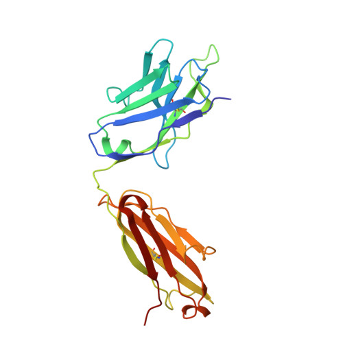
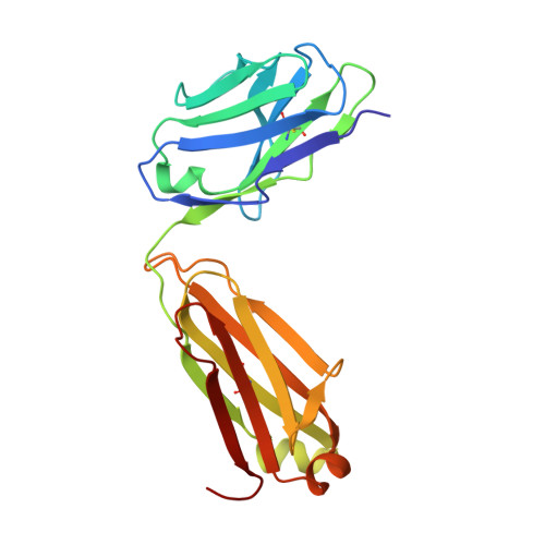
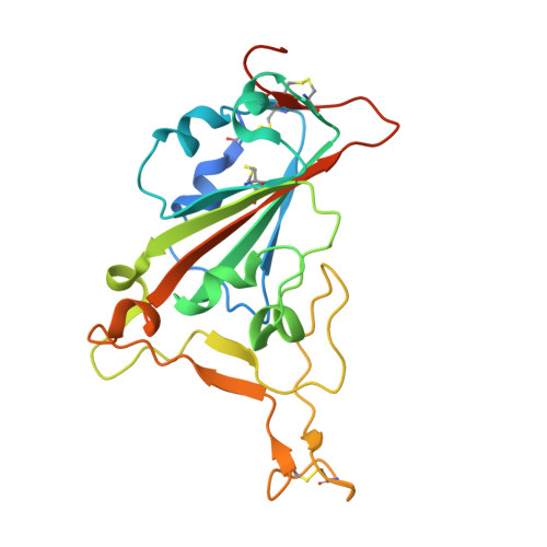
Crystal structure of SARS-CoV-2 Spike RBD in complex with MW01 Fab


Experimental Data Snapshot

  • Method: X-RAY DIFFRACTION
  • Resolution: 2.40 Å
  • R-Value Free: 0.230 
  • R-Value Work: 0.183 
  • R-Value Observed: 0.185 

Starting Models: experimental
View more details

wwPDB Validation   3D Report Full Report


Ligand Structure Quality Assessment 


This is version 1.3 of the entry. See complete history


Literature

Antibody-dependent enhancement (ADE) of SARS-CoV-2 pseudoviral infection requires Fc gamma RIIB and virus-antibody complex with bivalent interaction.

Wang, S.Wang, J.Yu, X.Jiang, W.Chen, S.Wang, R.Wang, M.Jiao, S.Yang, Y.Wang, W.Chen, H.Chen, B.Gu, C.Liu, C.Wang, A.Wang, M.Li, G.Guo, C.Liu, D.Zhang, J.Zhang, M.Wang, L.Gui, X.

(2022) Commun Biol 5: 262-262

  • DOI: https://doi.org/10.1038/s42003-022-03207-0
  • Primary Citation of Related Structures:  
    7DJZ, 7DK0

  • PubMed Abstract: 

    Understanding the underlying molecular mechanisms behind ADE of SARS-CoV-2 is critical for development of safe and effective therapies. Here, we report that two neutralizing mAbs, MW01 and MW05, could enhance the infection of SARS-CoV-2 pseudovirus on FcγRIIB-expressing B cells. X-ray crystal structure determination and S trimer-binding modeling showed that MW01 and MW05 could bind to RBDs in S trimer with both "up" and "down" states. While, the neutralizing mAb MW07, which has no ADE activity only binds to RBD in S trimer with "up" state. Monovalent MW01 and MW05 completely diminished the ADE activity compared with their bivalent counterparts. Moreover, both macropinocytosis and endocytosis are confirmed involving in ADE of SARS-CoV-2 pseudoviral infection. Blocking endosome transportation and lysosome acidification could inhibit the ADE activity mediated by MW05. Together, our results identified a novel ADE mechanism of SARS-CoV-2 pseudovirus in vitro, FcγRIIB-mediated uptake of SARS-CoV-2/mAb complex with bivalent interaction.


  • Organizational Affiliation

    Mabwell (Shanghai) Bioscience Co., Ltd, Shanghai, 201210, China.


Macromolecules
Find similar proteins by:  (by identity cutoff)  |  3D Structure
Entity ID: 1
MoleculeChains Sequence LengthOrganismDetailsImage
MW01 heavy chain226Homo sapiensMutation(s): 0 
Entity Groups  
Sequence Clusters30% Identity50% Identity70% Identity90% Identity95% Identity100% Identity
Sequence Annotations
Expand
  • Reference Sequence
Find similar proteins by:  (by identity cutoff)  |  3D Structure
Entity ID: 2
MoleculeChains Sequence LengthOrganismDetailsImage
MW01 light chain213Homo sapiensMutation(s): 0 
Entity Groups  
Sequence Clusters30% Identity50% Identity70% Identity90% Identity95% Identity100% Identity
Sequence Annotations
Expand
  • Reference Sequence
Find similar proteins by:  (by identity cutoff)  |  3D Structure
Entity ID: 3
MoleculeChains Sequence LengthOrganismDetailsImage
Spike protein S1223Severe acute respiratory syndrome coronavirus 2Mutation(s): 0 
Gene Names: S2
UniProt
Find proteins for P0DTC2 (Severe acute respiratory syndrome coronavirus 2)
Explore P0DTC2 
Go to UniProtKB:  P0DTC2
Entity Groups  
Sequence Clusters30% Identity50% Identity70% Identity90% Identity95% Identity100% Identity
UniProt GroupP0DTC2
Glycosylation
Glycosylation Sites: 1Go to GlyGen: P0DTC2-1
Sequence Annotations
Expand
  • Reference Sequence
Experimental Data & Validation

Experimental Data

  • Method: X-RAY DIFFRACTION
  • Resolution: 2.40 Å
  • R-Value Free: 0.230 
  • R-Value Work: 0.183 
  • R-Value Observed: 0.185 
  • Space Group: C 2 2 21
Unit Cell:
Length ( Å )Angle ( ˚ )
a = 65.622α = 90
b = 86.833β = 90
c = 293.278γ = 90
Software Package:
Software NamePurpose
HKL-2000data reduction
HKL-2000data scaling
PHENIXrefinement
PDB_EXTRACTdata extraction
PHASERphasing

Structure Validation

View Full Validation Report


Ligand Structure Quality Assessment 


Entry History 

Deposition Data

Revision History  (Full details and data files)

  • Version 1.0: 2021-06-09
    Type: Initial release
  • Version 1.1: 2022-06-22
    Changes: Database references
  • Version 1.2: 2023-11-29
    Changes: Data collection, Refinement description
  • Version 1.3: 2024-11-20
    Changes: Structure summary