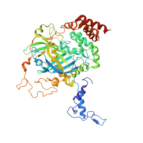
7DI8

Electron crystallographic structure of Catalase using a direct electron detector at 300 kV


Experimental Data Snapshot

  • Method: ELECTRON CRYSTALLOGRAPHY
  • Resolution: 3.20 Å
  • R-Value Free: 0.348 
  • R-Value Work: 0.309 
  • R-Value Observed: 0.311 

wwPDB Validation   3D Report Full Report


This is version 1.3 of the entry. See complete history


Literature

Protein and Organic-Molecular Crystallography With 300kV Electrons on a Direct Electron Detector.

Takaba, K.Maki-Yonekura, S.Inoue, S.Hasegawa, T.Yonekura, K.

(2020) Front Mol Biosci 7: 612226-612226

  • DOI: https://doi.org/10.3389/fmolb.2020.612226
  • Primary Citation of Related Structures:  
    7DI8

  • PubMed Abstract: 

    Electron 3D crystallography can reveal the atomic structure from undersized crystals of various samples owing to the strong scattering power of electrons. Here, a direct electron detector DE64 was tested for small and thin crystals of protein and an organic molecule using a JEOL CRYO ARM 300 electron microscope. The microscope is equipped with a cold-field emission gun operated at an accelerating voltage of 300 kV, quad condenser lenses for parallel illumination, an in-column energy filter, and a stable rotational goniometer stage. Rotational diffraction data were collected in an unsupervised manner from crystals of a heme-binding enzyme catalase and a representative organic semiconductor material Ph-BTBT-C10. The structures were determined by molecular replacement for catalase and by the direct method for Ph-BTBT-C10. The analyses demonstrate that the system works well for electron 3D crystallography of these molecules with less damaging, a smaller point spread, and less noise than using the conventional scintillator-coupled camera.


  • Organizational Affiliation

    Biostructural Mechanism Laboratory, RIKEN SPring-8 Center, Sayo, Japan.


Macromolecules
Find similar proteins by:  (by identity cutoff)  |  3D Structure
Entity ID: 1
MoleculeChains Sequence LengthOrganismDetailsImage
Catalase
A, B, C, D
527Bos taurusMutation(s): 0 
Gene Names: CAT
EC: 1.11.1.6
UniProt
Find proteins for P00432 (Bos taurus)
Explore P00432 
Go to UniProtKB:  P00432
Entity Groups  
Sequence Clusters30% Identity50% Identity70% Identity90% Identity95% Identity100% Identity
UniProt GroupP00432
Sequence Annotations
Expand
  • Reference Sequence
Small Molecules
Ligands 2 Unique
IDChains Name / Formula / InChI Key2D Diagram3D Interactions
NDP
Query on NDP

Download Ideal Coordinates CCD File 
F [auth A],
H [auth B],
J [auth C],
L [auth D]
NADPH DIHYDRO-NICOTINAMIDE-ADENINE-DINUCLEOTIDE PHOSPHATE
C21 H30 N7 O17 P3
ACFIXJIJDZMPPO-NNYOXOHSSA-N
HEM (Subject of Investigation/LOI)
Query on HEM

Download Ideal Coordinates CCD File 
E [auth A],
G [auth B],
I [auth C],
K [auth D]
PROTOPORPHYRIN IX CONTAINING FE
C34 H32 Fe N4 O4
KABFMIBPWCXCRK-RGGAHWMASA-L
Experimental Data & Validation

Experimental Data

  • Method: ELECTRON CRYSTALLOGRAPHY
  • Resolution: 3.20 Å
  • R-Value Free: 0.348 
  • R-Value Work: 0.309 
  • R-Value Observed: 0.311 
  • Space Group: P 21 21 21
Unit Cell:
Length ( Å )Angle ( ˚ )
a = 69.98α = 90
b = 174.02β = 90
c = 199.45γ = 90
Software Package:
Software NamePurpose
PHENIXrefinement
XSCALEdata scaling
PDB_EXTRACTdata extraction

Structure Validation

View Full Validation Report


Entry History & Funding Information

Deposition Data


Funding OrganizationLocationGrant Number
Japan Society for the Promotion of Science (JSPS)Japan24657111
Japan Agency for Medical Research and Development (AMED)Japan--

Revision History  (Full details and data files)

  • Version 1.0: 2020-12-09
    Type: Initial release
  • Version 1.1: 2021-01-27
    Changes: Database references
  • Version 1.2: 2021-02-03
    Changes: Database references
  • Version 1.3: 2024-03-27
    Changes: Data collection, Database references