7DHW | pdb_00007dhw

Crystal structure of myosin-XI motor domain in complex with ADP-ALF4


Experimental Data Snapshot

  • Method: X-RAY DIFFRACTION
  • Resolution: 2.84 Å
  • R-Value Free: 
    0.271 (Depositor), 0.274 (DCC) 
  • R-Value Work: 
    0.226 (Depositor), 0.230 (DCC) 
  • R-Value Observed: 
    0.231 (Depositor) 

Starting Model: experimental
View more details

wwPDB Validation 3D Report Full Report

Validation slider image for 7DHW

Ligand Structure Quality Assessment 


This is version 1.4 of the entry. See complete history

Literature

Discovery of ultrafast myosin, its amino acid sequence, and structural features.

Haraguchi, T.Tamanaha, M.Suzuki, K.Yoshimura, K.Imi, T.Tominaga, M.Sakayama, H.Nishiyama, T.Murata, T.Ito, K.

(2022) Proc Natl Acad Sci U S A 119

  • DOI: https://doi.org/10.1073/pnas.2120962119
  • Primary Citation Related Structures: 
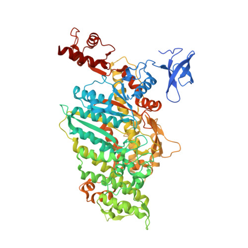
    7DHW

  • PubMed Abstract: 

    Cytoplasmic streaming with extremely high velocity (∼70 μm s -1 ) occurs in cells of the characean algae ( Chara ). Because cytoplasmic streaming is caused by myosin XI, it has been suggested that a myosin XI with a velocity of 70 μm s -1 , the fastest myosin measured so far, exists in Chara cells. However, the velocity of the previously cloned Chara corallina myosin XI ( Cc XI) was about 20 μm s -1 , one-third of the cytoplasmic streaming velocity in Chara Recently, the genome sequence of Chara braunii has been published, revealing that this alga has four myosin XI genes. We cloned these four myosin XI ( Cb XI-1, 2, 3, and 4) and measured their velocities. While the velocities of Cb XI-3 and Cb XI-4 motor domains (MDs) were similar to that of Cc XI MD, the velocities of Cb XI-1 and Cb XI-2 MDs were 3.2 times and 2.8 times faster than that of Cc XI MD, respectively. The velocity of chimeric Cb XI-1, a functional, full-length Cb XI-1 construct, was 60 μm s -1 These results suggest that Cb XI-1 and Cb XI-2 would be the main contributors to cytoplasmic streaming in Chara cells and show that these myosins are ultrafast myosins with a velocity 10 times faster than fast skeletal muscle myosins in animals. We also report an atomic structure (2.8-Å resolution) of myosin XI using X-ray crystallography. Based on this crystal structure and the recently published cryo-electron microscopy structure of acto-myosin XI at low resolution (4.3-Å), it appears that the actin-binding region contributes to the fast movement of Chara myosin XI. Mutation experiments of actin-binding surface loops support this hypothesis.


  • Organizational Affiliation
    • Department of Biology, Graduate School of Science, Chiba University, Chiba 263-8522, Japan.

Macromolecule Content 

  • Total Structure Weight: 88.42 kDa 
  • Atom Count: 5,778 
  • Modeled Residue Count: 726 
  • Deposited Residue Count: 771 
  • Unique protein chains: 1

Macromolecules

Find similar proteins by:|  3D Structure
Entity ID: 1
MoleculeChains  Sequence LengthOrganismDetailsImage
Motor domain of myosin771Arabidopsis thalianaMutation(s): 0 
Gene Names: XI-2MYA2At5g43900F6B6.4
UniProt
Find proteins for Q9LKB9 (Arabidopsis thaliana)
Explore Q9LKB9 
Go to UniProtKB:  Q9LKB9
Entity Groups
Sequence Clusters30% Identity50% Identity70% Identity90% Identity95% Identity100% Identity
UniProt GroupQ9LKB9
Sequence Annotations
Expand
Reference Sequence

Small Molecules

Ligands 4 Unique
IDChains Name / Formula / InChI Key2D Diagram3D Interactions
ADP
(Subject of Investigation/LOI)

Query on ADP


Download:Ideal Coordinates CCD File
E [auth A]ADENOSINE-5'-DIPHOSPHATE
C10 H15 N5 O10 P2
XTWYTFMLZFPYCI-KQYNXXCUSA-N
ALF

Query on ALF


Download:Ideal Coordinates CCD File
D [auth A]TETRAFLUOROALUMINATE ION
Al F4
UYOMQIYKOOHAMK-UHFFFAOYSA-J
GOL

Query on GOL


Download:Ideal Coordinates CCD File
F [auth A],
G [auth A],
H [auth A],
I [auth A]
GLYCEROL
C3 H8 O3
PEDCQBHIVMGVHV-UHFFFAOYSA-N
MG

Query on MG


Download:Ideal Coordinates CCD File
B [auth A],
C [auth A]
MAGNESIUM ION
Mg
JLVVSXFLKOJNIY-UHFFFAOYSA-N

Experimental Data & Validation

Experimental Data

  • Method: X-RAY DIFFRACTION
  • Resolution: 2.84 Å
  • R-Value Free:  0.271 (Depositor), 0.274 (DCC) 
  • R-Value Work:  0.226 (Depositor), 0.230 (DCC) 
  • R-Value Observed: 0.231 (Depositor) 
Space Group: P 21 21 21
Unit Cell:
Length ( Å )Angle ( ˚ )
a = 58.277α = 90
b = 73.725β = 90
c = 187.957γ = 90
Software Package:
Software NamePurpose
PHENIXrefinement
XDSdata reduction
XDSdata scaling
PHASERphasing
Cootmodel building

Structure Validation

View Full Validation Report


Ligand Structure Quality Assessment 


Entry History 

Deposition Data

Revision History  (Full details and data files)

  • Version 1.0: 2021-05-19
    Type: Initial release
  • Version 1.1: 2021-10-13
    Changes: Database references, Structure summary
  • Version 1.2: 2022-02-23
    Changes: Database references
  • Version 1.3: 2022-03-02
    Changes: Database references
  • Version 1.4: 2023-11-29
    Changes: Data collection, Refinement description