


The co-crystal structure of DYRK2 with a small molecule inhibitor 5
Wei, T., Xiao, J.To be published.
Experimental Data Snapshot
Starting Model: experimental
View more details
Entity ID: 1 | |||||
|---|---|---|---|---|---|
| Molecule | Chains | Sequence Length | Organism | Details | Image |
| Dual specificity tyrosine-phosphorylation-regulated kinase 2 | 326 | Homo sapiens | Mutation(s): 0 Gene Names: DYRK2 EC: 2.7.12.1 | 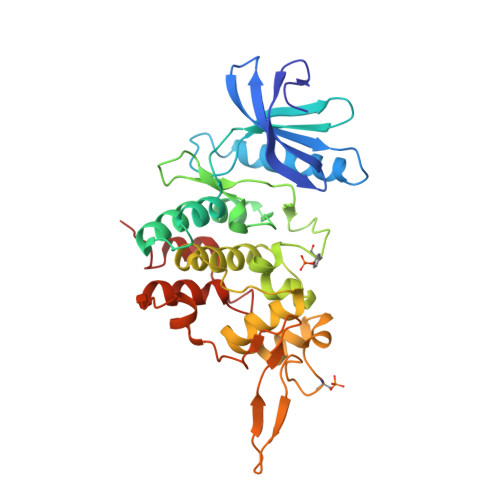 | |
UniProt & NIH Common Fund Data Resources | |||||
PHAROS: Q92630 GTEx: ENSG00000127334 | |||||
Entity Groups | |||||
| Sequence Clusters | 30% Identity50% Identity70% Identity90% Identity95% Identity100% Identity | ||||
| UniProt Group | Q92630 | ||||
Sequence AnnotationsExpand | |||||
Reference Sequence | |||||
| Ligands 1 Unique | |||||
|---|---|---|---|---|---|
| ID | Chains | Name / Formula / InChI Key | 2D Diagram | 3D Interactions | |
| H7C (Subject of Investigation/LOI) Download:Ideal Coordinates CCD File | B [auth A] | 2,7-dimethoxy-9-piperidin-4-ylsulfanyl-acridine C20 H22 N2 O2 S ZXEZPSYSBIFZGE-UHFFFAOYSA-N |  | ||
| Modified Residues 2 Unique | |||||
|---|---|---|---|---|---|
| ID | Chains | Type | Formula | 2D Diagram | Parent |
| PTR Query on PTR | A | L-PEPTIDE LINKING | C9 H12 N O6 P |  | TYR |
| SEP Query on SEP | A | L-PEPTIDE LINKING | C3 H8 N O6 P |  | SER |
| Length ( Å ) | Angle ( ˚ ) |
|---|---|
| a = 64.56 | α = 90 |
| b = 128.83 | β = 90 |
| c = 132.45 | γ = 90 |
| Software Name | Purpose |
|---|---|
| PHENIX | refinement |
| XDS | data reduction |
| PDB_EXTRACT | data extraction |
| Aimless | data scaling |
| PHASER | phasing |
| Funding Organization | Location | Grant Number |
|---|---|---|
| National Science Foundation (NSF, China) | China | -- |


