A backbone-centred energy function of neural networks for protein design.
Huang, B., Xu, Y., Hu, X., Liu, Y., Liao, S., Zhang, J., Huang, C., Hong, J., Chen, Q., Liu, H.(2022) Nature 602: 523-528
- PubMed: 35140398 Search on PubMed
- DOI: https://doi.org/10.1038/s41586-021-04383-5
- Primary Citation Related Structures:
7DGU, 7DGW, 7DGY, 7DKK, 7DKO, 7DMF, 7FBB, 7FBC, 7FBD - PubMed Abstract:
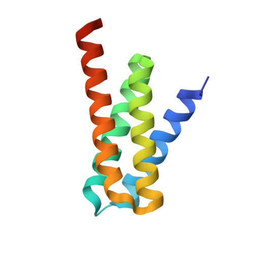
A protein backbone structure is designable if a substantial number of amino acid sequences exist that autonomously fold into it 1,2 . It has been suggested that the designability of backbones is governed mainly by side chain-independent or side chain type-insensitive molecular interactions 3-5 , indicating an approach for designing new backbones (ready for amino acid selection) based on continuous sampling and optimization of the backbone-centred energy surface. However, a sufficiently comprehensive and precise energy function has yet to be established for this purpose. Here we show that this goal is met by a statistical model named SCUBA (for Side Chain-Unknown Backbone Arrangement) that uses neural network-form energy terms. These terms are learned with a two-step approach that comprises kernel density estimation followed by neural network training and can analytically represent multidimensional, high-order correlations in known protein structures. We report the crystal structures of nine de novo proteins whose backbones were designed to high precision using SCUBA, four of which have novel, non-natural overall architectures. By eschewing use of fragments from existing protein structures, SCUBA-driven structure design facilitates far-reaching exploration of the designable backbone space, thus extending the novelty and diversity of the proteins amenable to de novo design.
- MOE Key Laboratory for Membraneless Organelles and Cellular Dynamics, Hefei National Laboratory for Physical Sciences at the Microscale, School of Life Sciences, Division of Life Sciences and Medicine, University of Science and Technology of China, Hefei, China.
Organizational Affiliation: