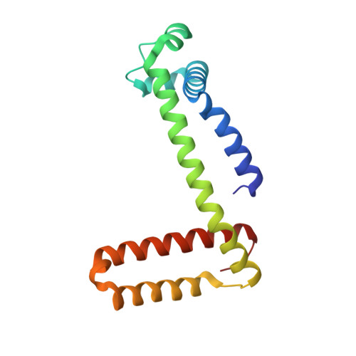
7DGN

The Co-bound dimeric structure of K79H/G80A/H81A myoglobin


Experimental Data Snapshot

  • Method: X-RAY DIFFRACTION
  • Resolution: 2.35 Å
  • R-Value Free: 0.269 
  • R-Value Work: 0.202 
  • R-Value Observed: 0.205 

Starting Model: experimental
View more details

wwPDB Validation   3D Report Full Report


Ligand Structure Quality Assessment 


This is version 1.2 of the entry. See complete history


Literature

Rational design of metal-binding sites in domain-swapped myoglobin dimers.

Nagao, S.Idomoto, A.Shibata, N.Higuchi, Y.Hirota, S.

(2021) J Inorg Biochem 217: 111374-111374

  • DOI: https://doi.org/10.1016/j.jinorgbio.2021.111374
  • Primary Citation of Related Structures:  
    7DGJ, 7DGK, 7DGL, 7DGM, 7DGN, 7DGO

  • PubMed Abstract: 

    The metal active site is precisely designed in metalloproteins. Here we applied 3D domain swapping, a phenomenon in which a partial protein structure is exchanged between molecules, to introduce metal sites in proteins. We designed multiple metal-binding sites specific to domain-swapped myoglobin (Mb) with His mutation. Stable dimeric Mbs with metal-binding sites were obtained by shifting the His position and introducing two Ala residues in the hinge region (K78H/G80A/H82A and K79H/G80A/H81A Mbs). The absorption and circular dichroism spectra of the monomer and dimer of K78H/G80A/H82A and K79H/G80A/H81A Mbs were similar to the corresponding spectra, respectively, of wild-type Mb. No negative peak due to dimer-to-monomer dissociation was observed below the denaturation temperature in the differential scanning calorimetry thermograms of K78H/G80A/H82A and K79H/G80A/H81A Mbs, whereas the dimer dissociates into monomers at 68 °C for wild-type Mb. These results show that the two mutants were stable in the dimer state. Metal ions bound to the metal-binding sites containing the introduced His in the domain-swapped Mb dimers. Co 2+ -bound and Ni 2+ -bound K78H/G80A/H82A Mb exhibited octahedral metal-coordination structures, where His78, His81, Glu85, and three H 2 O/OH - molecules coordinated to the metal ion. On the other hand, Co 2+ -bound and Zn 2+ -bound K79H/G80A/H81A Mb exhibited tetrahedral metal-coordination structures, where His79, His82, Asp141, and a H 2 O/OH - molecule coordinated to the metal ion. The Co 2+ -bound site exists deep inside the protein in the K79H/G80A/H81A Mb dimer, which may allow the unique tetrahedral coordination for the Co 2+ ion. These results show that we can utilize domain swapping to construct artificial metalloproteins.


  • Organizational Affiliation

    Division of Materials Science, Graduate School of Science and Technology, Nara Institute of Science and Technology, 8916-5 Takayama, Ikoma, Nara 630-0192, Japan. Electronic address: s-nagao@sci.u-hyogo.ac.jp.


Macromolecules
Find similar proteins by:  (by identity cutoff)  |  3D Structure
Entity ID: 1
MoleculeChains Sequence LengthOrganismDetailsImage
Myoglobin
A, B
153Equus caballusMutation(s): 3 
Gene Names: MB
EC: 1.11.1 (UniProt), 1.7 (UniProt)
UniProt
Find proteins for P68082 (Equus caballus)
Explore P68082 
Go to UniProtKB:  P68082
Entity Groups  
Sequence Clusters30% Identity50% Identity70% Identity90% Identity95% Identity100% Identity
UniProt GroupP68082
Sequence Annotations
Expand
  • Reference Sequence
Small Molecules
Ligands 3 Unique
IDChains Name / Formula / InChI Key2D Diagram3D Interactions
HEM (Subject of Investigation/LOI)
Query on HEM

Download Ideal Coordinates CCD File 
C [auth A],
G [auth B]
PROTOPORPHYRIN IX CONTAINING FE
C34 H32 Fe N4 O4
KABFMIBPWCXCRK-RGGAHWMASA-L
CO (Subject of Investigation/LOI)
Query on CO

Download Ideal Coordinates CCD File 
E [auth A],
H [auth B]
COBALT (II) ION
Co
XLJKHNWPARRRJB-UHFFFAOYSA-N
O
Query on O

Download Ideal Coordinates CCD File 
D [auth A],
F [auth B]
OXYGEN ATOM
O
XLYOFNOQVPJJNP-UHFFFAOYSA-N
Experimental Data & Validation

Experimental Data

  • Method: X-RAY DIFFRACTION
  • Resolution: 2.35 Å
  • R-Value Free: 0.269 
  • R-Value Work: 0.202 
  • R-Value Observed: 0.205 
  • Space Group: P 21 21 21
Unit Cell:
Length ( Å )Angle ( ˚ )
a = 56.966α = 90
b = 63.463β = 90
c = 83.003γ = 90
Software Package:
Software NamePurpose
REFMACrefinement
XDSdata reduction
SCALAdata scaling
MOLREPphasing

Structure Validation

View Full Validation Report


Ligand Structure Quality Assessment 


Entry History & Funding Information

Deposition Data


Funding OrganizationLocationGrant Number
Japan Society for the Promotion of Science (JSPS)JapanJP16K17935
Japan Society for the Promotion of Science (JSPS)JapanJP19K05695
Japan Society for the Promotion of Science (JSPS)JapanJP26288080

Revision History  (Full details and data files)

  • Version 1.0: 2021-02-17
    Type: Initial release
  • Version 1.1: 2021-03-03
    Changes: Database references
  • Version 1.2: 2023-11-29
    Changes: Data collection, Database references, Refinement description