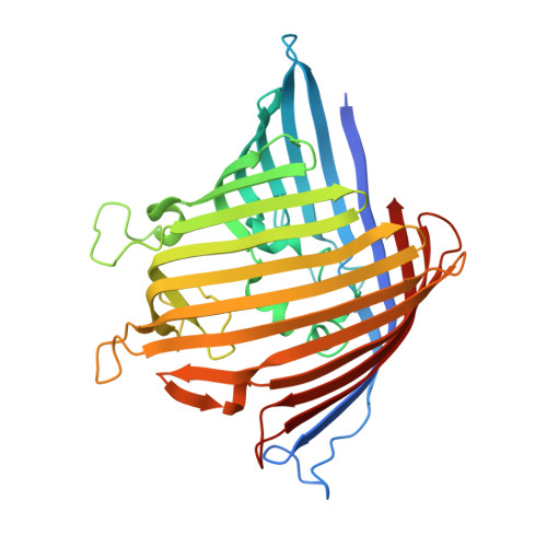
An antibiotic-resistance conferring mutation in a neisserial porin: Structure, ion flux, and ampicillin binding.
Bartsch, A., Ives, C.M., Kattner, C., Pein, F., Diehn, M., Tanabe, M., Munk, A., Zachariae, U., Steinem, C., Llabres, S.(2021) Biochim Biophys Acta Biomembr 1863: 183601-183601
- PubMed: 33675718
- DOI: https://doi.org/10.1016/j.bbamem.2021.183601
- Primary Citation of Related Structures:
7DE8 - PubMed Abstract:
Gram-negative bacteria cause the majority of highly drug-resistant bacterial infections. To cross the outer membrane of the complex Gram-negative cell envelope, antibiotics permeate through porins, trimeric channel proteins that enable the exchange of small polar molecules. Mutations in porins contribute to the development of drug-resistant phenotypes. In this work, we show that a single point mutation in the porin PorB from Neisseria meningitidis, the causative agent of bacterial meningitis, can strongly affect the binding and permeation of beta-lactam antibiotics. Using X-ray crystallography, high-resolution electrophysiology, atomistic biomolecular simulation, and liposome swelling experiments, we demonstrate differences in drug binding affinity, ion selectivity and drug permeability of PorB. Our work further reveals distinct interactions between the transversal electric field in the porin eyelet and the zwitterionic drugs, which manifest themselves under applied electric fields in electrophysiology and are altered by the mutation. These observations may apply more broadly to drug-porin interactions in other channels. Our results improve the molecular understanding of porin-based drug-resistance in Gram-negative bacteria.
- Institute of Organic and Biomolecular Chemistry, University of Göttingen, Tammannstraße 2, 37077 Göttingen, Germany.
Organizational Affiliation:


