7DBJ | pdb_00007dbj

Crystal structure of human LDHB in complex with NADH, oxamate, and AXKO-0046


Experimental Data Snapshot

  • Method: X-RAY DIFFRACTION
  • Resolution: 1.55 Å
  • R-Value Free: 
    0.192 (Depositor), 0.197 (DCC) 
  • R-Value Work: 
    0.170 (Depositor), 0.179 (DCC) 

Starting Model: experimental
View more details

wwPDB Validation 3D Report Full Report

Validation slider image for 7DBJ

Ligand Structure Quality Assessment 


This is version 1.3 of the entry. See complete history

Literature

Identification of the first highly selective inhibitor of human lactate dehydrogenase B.

Shibata, S.Sogabe, S.Miwa, M.Fujimoto, T.Takakura, N.Naotsuka, A.Kitamura, S.Kawamoto, T.Soga, T.

(2021) Sci Rep 11: 21353-21353

  • DOI: https://doi.org/10.1038/s41598-021-00820-7
  • Primary Citation Related Structures: 
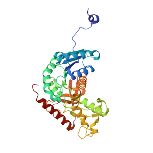
    7DBJ, 7DBK

  • PubMed Abstract: 

    Lactate dehydrogenase (LDH) catalyses the conversion of pyruvate to lactate and NADH to NAD + ; it has two isoforms, LDHA and LDHB. LDHA is a promising target for cancer therapy, whereas LDHB is necessary for basal autophagy and cancer cell proliferation in oxidative and glycolytic cancer cells. To the best of our knowledge, selective inhibitors for LDHB have not yet been reported. Here, we developed a high-throughput mass spectrometry screening system using an LDHB enzyme assay by detecting NADH and NAD + . As a result, we identified a small-molecule LDHB selective inhibitor AXKO-0046, an indole derivative. This compound exhibited uncompetitive LDHB inhibition (EC 50  = 42 nM). X-ray crystallography revealed that AXKO-0046 bound to the potential allosteric site away from the LDHB catalytic active site, suggesting that targeting the tetramerisation interface of the two dimers is critical for the enzymatic activity. AXKO-0046 and its derivatives can be used to validate LDHB-associated pathways in cancer metabolism.


  • Organizational Affiliation
    • Discovery Biology, Discovery Science, Axcelead Drug Discovery Partners, Inc., 2-26-1 Muraoka-Higashi, Fujisawa, Kanagawa, Japan. sachio.shibata@axcelead.com.

Macromolecule Content 

  • Total Structure Weight: 150.4 kDa 
  • Atom Count: 11,302 
  • Modeled Residue Count: 1,332 
  • Deposited Residue Count: 1,332 
  • Unique protein chains: 1

Macromolecules

Find similar proteins by:|  3D Structure
Entity ID: 1
MoleculeChains  Sequence LengthOrganismDetailsImage
L-lactate dehydrogenase B chain
A, B, C, D
333Homo sapiensMutation(s): 0 
Gene Names: LDHB
EC: 1.1.1.27
UniProt & NIH Common Fund Data Resources
Find proteins for P07195 (Homo sapiens)
Explore P07195 
Go to UniProtKB:  P07195
PHAROS:  P07195
GTEx:  ENSG00000111716 
Entity Groups
Sequence Clusters30% Identity50% Identity70% Identity90% Identity95% Identity100% Identity
UniProt GroupP07195
Sequence Annotations
Expand
Reference Sequence

Small Molecules

Ligands 4 Unique
IDChains Name / Formula / InChI Key2D Diagram3D Interactions
NAI

Query on NAI


Download:Ideal Coordinates CCD File
E [auth A],
H [auth B],
L [auth C],
Q [auth D]
1,4-DIHYDRONICOTINAMIDE ADENINE DINUCLEOTIDE
C21 H29 N7 O14 P2
BOPGDPNILDQYTO-NNYOXOHSSA-N
H1U

Query on H1U


Download:Ideal Coordinates CCD File
K [auth C],
P [auth D]
N-[[3-[2-[(phenylmethyl)amino]ethyl]-1H-indol-2-yl]methyl]cycloheptanamine
C25 H33 N3
VJTXDVYRAXQJAK-UHFFFAOYSA-N
OXM

Query on OXM


Download:Ideal Coordinates CCD File
F [auth A],
I [auth B],
M [auth C],
R [auth D]
OXAMIC ACID
C2 H3 N O3
SOWBFZRMHSNYGE-UHFFFAOYSA-N
EDO

Query on EDO


Download:Ideal Coordinates CCD File
G [auth A]
J [auth B]
N [auth C]
O [auth C]
S [auth D]
G [auth A],
J [auth B],
N [auth C],
O [auth C],
S [auth D],
T [auth D],
U [auth D]
1,2-ETHANEDIOL
C2 H6 O2
LYCAIKOWRPUZTN-UHFFFAOYSA-N
Binding Affinity Annotations 
IDSourceBinding Affinity
OXM BindingDB:  7DBJ Ki: 9.44e+4 (nM) from 1 assay(s)
IC50: min: 3.38e+4, max: 1.23e+5 (nM) from 3 assay(s)

Experimental Data & Validation

Experimental Data

  • Method: X-RAY DIFFRACTION
  • Resolution: 1.55 Å
  • R-Value Free:  0.192 (Depositor), 0.197 (DCC) 
  • R-Value Work:  0.170 (Depositor), 0.179 (DCC) 
Space Group: P 1 21 1
Unit Cell:
Length ( Å )Angle ( ˚ )
a = 59.389α = 90
b = 137.577β = 109.264
c = 84.909γ = 90
Software Package:
Software NamePurpose
REFMACrefinement
HKL-2000data reduction
SCALEPACKdata scaling
PHASERphasing

Structure Validation

View Full Validation Report


Ligand Structure Quality Assessment 


Entry History 

Deposition Data

Revision History  (Full details and data files)

  • Version 1.0: 2021-10-20
    Type: Initial release
  • Version 1.1: 2021-11-10
    Changes: Database references
  • Version 1.2: 2022-02-16
    Changes: Database references
  • Version 1.3: 2023-11-29
    Changes: Data collection, Derived calculations, Refinement description